Review
. 2021 Oct 16;19(1):325.
doi: 10.1186/s12951-021-01074-1.

Fenton/Fenton-like metal-based nanomaterials combine with oxidase for synergistic tumor therapy

Affiliations
Review

Fenton/Fenton-like metal-based nanomaterials combine with oxidase for synergistic tumor therapy

Wei Cao et al. J Nanobiotechnology. .

Abstract

Chemodynamic therapy (CDT) catalyzed by transition metal and starvation therapy catalyzed by intracellular metabolite oxidases are both classic tumor treatments based on nanocatalysts. CDT monotherapy has limitations including low catalytic efficiency of metal ions and insufficient endogenous hydrogen peroxide (H2O2). Also, single starvation therapy shows limited ability on resisting tumors. The "metal-oxidase" cascade catalytic system is to introduce intracellular metabolite oxidases into the metal-based nanoplatform, which perfectly solves the shortcomings of the above-mentioned monotherapiesIn this system, oxidases can not only consume tumor nutrients to produce a "starvation effect", but also provide CDT with sufficient H2O2 and a suitable acidic environment, which further promote synergy between CDT and starvation therapy, leading to enhanced antitumor effects. More importantly, the "metal-oxidase" system can be combined with other antitumor therapies (such as photothermal therapy, hypoxia-activated drug therapy, chemotherapy, and immunotherapy) to maximize their antitumor effects. In addition, both metal-based nanoparticles and oxidases can activate tumor immunity through multiple pathways, so the combination of the "metal-oxidase" system with immunotherapy has a powerful synergistic effect. This article firstly introduced the metals which induce CDT and the oxidases which induce starvation therapy and then described the "metal-oxidase" cascade catalytic system in detail. Moreover, we highlight the application of the "metal-oxidase" system in combination with numerous antitumor therapies, especially in combination with immunotherapy, expecting to provide new ideas for tumor treatment.

Keywords: Chemodynamic therapy; Fenton reaction; Immunotherapy; Metabolite oxidase; Tumor synergistic therapy.


Conflict of interest statement

The authors declare that they have no competing interests.

Figures
Scheme 1
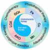
Scheme 1
Schematic diagram of the synergistic anti-tumor effect of the "metal-oxidase" cascade catalytic system and the combination of this system with other anti-tumor therapies
Fig. 1
Fig. 1
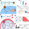
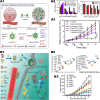
Iron-mediated Fenton reaction triggers CDT. A1 Preparation of AFeNPs. A2 The optimal value for AFeNPs catalyzed hydrogen peroxide decomposition was pH = 5.4. A3 Changes in relative volume of tumors after different treatments. Reproduced with permission from Ref [28], copyright © 2016 Wiley–VCH. B1 Schematic diagram of tumor therapeutic performance of biodegradable rFeOx-HMSN nanoparticles. B2 (a) Body weight changes of 4T1 bearing mice after different treatments. Relative tumor volumes of 4T1-bearing mice administrated with varied doses of PEG/rFeOx-HMSN nanocatalyst (b) intratumorally and (c) intravenously. Digital photographs of dissected tumors after the therapeutic process from (d) intratumoral and (e) intravenous groups. Reproduced with permission from Ref [29], copyright © 2018 Elsevier. C1 Schematic of CuS-Fe boosting transformation of Fe (III) into Fe (II) for highly improving CDT. C2 GSH level in HeLa cells after difffferent treatments; Cell viability in HeLa cells and NIH3T3 cells after different treatments. C3 Changes in body weight of mice and tumor volume during different treatments. Reproduced with permission from Ref [30], copyright © 2019 American Chemical Society
Fig. 2
Fig. 2
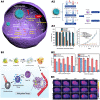
Copper element mediates Fenton reaction to trigger CDT. A1 Schematic of the Cu-Cys NPs synthetic process and the copper-containing nanoformulation mediated CDT. A2 GSH/GSSG ratio in MCF-7R cells before and after treatment with 100 μg/mL of Cu-Cys NPs. A3 Changes in body weight of mice and tumor volume during different treatments. Reproduced with permission from Ref [34], copyright © 2018 American Chemical Society. B1 Formation of CP nanodots for H2O2 self-supplying CDT. B2 Flow cytometry analysis of apoptosis in U87MG cells treated with CP nanodots for 12 h and CDT potency of CP nanodots after 24 h of incubation. B3 Biodistribution of Cu in major organs and tumor of U87MG tumor-bearing mice at 24 h post i.v. injection with CP nanodots and changes in body weight of mice and tumor volume during different treatments. Reproduced with permission from Ref [36], copyright © 2019 American Chemical Society
Fig. 3
Fig. 3
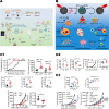
Manganese mediates the Fenton reaction to trigger CDT. A1 Schematic illustrating the application of MS@MnO2 NPs for MRI-monitored chemo-chemodynamic combination therapy. A2 Viability of U87MG cells after 24 h of different treatments. And viability of U87MG cells after 24 h of different treatments. And H&E-stained images of tumour sections from different groups. Reproduced with permission from Ref [39], copyright © 2018 WILEY–VCH. B1 Illustration of the synthetic process of MCDION-Se. And the cascade reaction of MCDION-Se in the intracellular environment. B2 The higher the degree of acidity the stronger the catalytic reaction. B3 Relevant in vitro experiments and in vivo experiments demonstrated the efficacy and safety of MCDION-Se. Reproduced with permission from Ref [40], copyright © 2019 Elsevier. C1 Schematic diagram and efficacy of LCN@Mn-BA nanoparticles for anti-tumor. C2 In vivo experiments in mice demonstrated LCN@Mn-BA can effectively inhibit tumor growth with good biosafety. Reproduced with permission from Ref [38], copyright © 2020 Elsevier
Fig. 4
Fig. 4
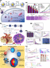
Glucose oxidase-mediated starvation therapy. A1 Schematic illustration of the mCGP nanoparticles for cancer targeting starvation therapy and PDT. A2 The dark and light toxicities of mCGP against 4T1 cells by MTT assay under 21% O2 or 2% O2. A3 The anticancer efficiency of mCGP in vivo. Reproduced with permission from Ref [56], copyright © 2017, American Chemical Society. B1 Illustration of GOx-induced starvation for enhanced low-temperature PTT in a hypoxic TME. B2 Representative Western blotting of HSP 90 and HSP 70 expression under different conditions. B3 In vivo experiments in mice, the nanoparticles were able to effectively inhibit tumors and prolong mouse survival time. Reproduced with permission from Ref [53], copyright © 2018, American Chemical Society
Fig. 5
Fig. 5
Lactate oxidase-mediated starvation therapy. A1 Schematic diagram of tumor-targeted CPGL micelles as the combination of starving and catalytic therapy basing on the generation of highly toxic ▪OH. A2 In vitro cytotoxicity of B16 cells after treated with CPGL micelles (0–0.9 μg/mL) for 24 h under different conditions (pH 7.4 and pH 6.0). A3 Monitoring body weight of mice every 2 days. And tumor volumes of B16 bearing mice after injections intravenously. The yellow arrow showed the injection time. Reproduced with permission from Ref [73], copyright © 2020, American Chemical Society. B1 Illustration of lactate-depletion-enabled TME adjustment and combinational cancer treatment strategy. B2 Tumor growth curves of mice after different treatments. And lactate concentration in 4T1 tumors at 48 h post-injections. B3 Numbers of blood vessels/field and percentages of VEGF-stained area were analyzed in tumor sections. Reproduced with permission from Ref [50], copyright © 2020 WILEY–VCH
Fig. 6
Fig. 6
"Iron-GOx" synergistic anti-tumor effect. A1 Fabrication and catalytic-therapeutic schematics of sequential GFD NCs. A2 In vivo catalytic-therapeutic performance of GOx-Fe3O4@DMSN against 4T1 and U87 tumor xenografts. Reproduced with permission from Ref [59], copyright © 2017 Springer Nature. B1 Schematic illustration of the preparation of NMIL-100@GOx@C and the cascade processes for cancer therapy. B2 GSH triggered Fe2+ release from NMIL-100. And the pH value changes of NMIL-100@GOx@C solution with or without glucose. B3 Average tumor weights and tumor growth curve in different treatment groups. Reproduced with permission from Ref [86], copyright © 2020, American Chemical Society. C1 Synthetic procedure and corresponding therapeutic principle of GOx-FeIIITA nanocomposites. C2 Viability of MCF-7 cells after the incubation with different concentrations of FeIIITA or GOx-FeIIITA nanocomposites. And cell apoptosis of MCF-7 cells after the incubation with different agents. C3 Average body weight and relative tumor volume in different groups subjected to various treatments. Reproduced with permission from Ref [88], copyright © 2019 IOP Publishing
Fig. 7
Fig. 7
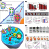
Synergistic effect of "copper-GOx" cascade catalytic system in tumor therapy. A1 Scheme of synthetic process and therapeutic mechanism of HMSN-Cu-GOD. A2 Cytotoxicity of HMSN-Cu-GOD treatment for MCF-7 cells after 24 h of incubation. A3 Body weight and tumor volumes change during 15 days of observation periods in different groups subjected to various treatments. Reproduced with permission from Ref [89], copyright © 2020 Elsevier. B1 Schematic illustration of the main synthesis procedures and antitumor mechanism of PCN-224(Cu)-GOx@MnO2 nMOFs. B2 Cell viability with different treatment. B3 In vivo anticancer efficacy of PCN-224(Cu)-GOx@MnO2 nMOFs. Reproduced with permission from Ref [90], copyright © 2020, American Chemical Society
Fig. 8
Fig. 8
The synergistic anti-tumor effect of " iron-LOX " and "iron-AAO". A1 The tandem biological–chemical catalytic reactions for effective catalytic tumor treatment based on the characteristic of TME. A2 Cell rescue profiles of 4T1 cells’ cytotoxicity induced by LFZ NPs (10 μg/mL) with different concentrations of l-ascorbic acid. A3 The relative tumor volume and the body weight under different treatments. Reproduced with permission from Ref [91], copyright © 2020, American Chemical Society. B1 Schematic illustration for the functioning mechanism of M@AAO@HFe–TA nanocapsules. B2 Protein expression of Bcl-2/Bax/Cyt C/caspase 3 mitochondrial apoptotic pathway in 4T1 cells under different treatments. And schematic illustration of in vitro antitumor mechanism of M@AAO@HFe–TA nanocapsules. B3 Body weight and tumor volume in different mice groups during the 14 days of treatment. Reproduced with permission from Ref [94], copyright © 2020, Royal Society of Chemistry
Fig. 9
Fig. 9
The synergistic anti-tumor system of "metal-oxidase" system combined with chemotherapy. A1 Diagram of how Fe/G@R-NRs is synthesized and how it is in an acidic environment at disassembly further and induces multiple antitumor responses. A2 Cytotoxicity against A549 cells at various CPT-equivalent concentrations. A3 Time-dependent A549 tumor growth profiles. The arrows indicate the injection time. Mice body weight change over the time during the treatment. The survival curve of A549 tumor-bearing mice after various treatments. Reproduced with permission from Ref [103], copyright © 2019, American Chemical Society. (B) B1 schematic illustration of GOx-MnCaP-DOX applied for MRI-monitored cooperative cancer therapy. B2 Average body weight and relative tumor volume in different groups subjected to various treatments. And H&E staining images of tumor tissues excised from the mice at day 14. Reproduced with permission from Ref [105], copyright © 2019, American Chemical Society
Fig. 10
Fig. 10
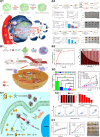
"Metal-oxidase" system combined with hypoxia activation therapy synergistic anti-tumor system. A1 Scheme illustrating the design of combining starvation and hypoxia-activated therapy by co-delivery of liposome-GOx and liposome-AQ4N into tumors. A2 Relative viabilities of 4T1 cells after 24 h incubated with different concentrations of glucose and liposome-GOx. And relative viabilities of 4T1 cells after 48 h incubation with different concentrations of liposome-AQ4N under the normoxia or hypoxia culture condition. A3 Tumor growth curves of mice after various different treatments indicated. Reproduced with permission from Ref [48], copyright © 2018 Elsevier. B1 Schematic diagram showing the fabrication of HGTFT nanoreactors and their applications for starvation therapy, CDT, and chemotherapy. B2 Tumor growth inhibition rates of different formulations. And apoptosis/necrosis rates determined by the TUNEL assay. B3 Tumor growth profiles of the mice in the different groups. Reproduced with permission from Ref [107], copyright © 2020 WILEY–VCH
Fig. 11
Fig. 11
Synergistic anti-tumor system of "metal-oxidase" system combined with photothermal therapy. A1 Schematic illustration of TME-based Fe(II)-PDA-GOD nanosystems for efficient cancer therapy by combining glucose degradation, Fenton reaction and photothermal therapy. A2 Schematic illustration of photothermal effect and catalytic performance of Fe(II) -PDA-GOD NPs and their in vitro antitumor effect. A3 The antitumor effects of nanoparticles in vivo and in vitro under different treatments. Reproduced with permission from Ref [109], copyright © 2019, American Chemical Society. B1 Schematic illustration of the construction of CuS-PGH NMs for multi-gradient antitumor therapy. B2 Changes of 4T1 cell viability after different treatments. B3 The real-time thermal images recorded using an IR thermal camera. Reproduced with permission from Ref [110], copyright © 2020, Royal Society of Chemistry
Fig. 12
Fig. 12
Metal-based nanoparticles mediate immune activation. A Schematic illustration of the FePt/BP–PEI–FA NCs enhanced immunotherapy. Reproduced with permission from Ref [16], copyright © 2020, Royal Society of Chemistry. B Antitumor effect of Cu-PPT nanoparticles combined with immune checkpoint inhibitors. Reproduced with permission from Ref [116], copyright © 2020, American Chemical Society. C1 Mn is essential for immune responses against tumors. Mn2+ stimulates CD8+ T cells activation and promotes DCs maturation and antigen presentation. C2 Mn2+ mediated cGAS-STING pathway to activate tumor immunity. C3 Mn2+ boosts antitumor immunotherapy in mice. Reproduced with permission from Ref [41], copyright Copyright © 2020, Springer Nature
Fig. 13
Fig. 13
Oxidases can relieve the immunosuppression of TME in many ways. A Glucose oxidase mediated immune activation. A1 Schematic illustration of anti-tumor immune response and enhanced anti-PD-1 immunotherapy induced by CMSN-GOx. A2 Tumor volume and survival curves in the mice after different treatments. A3 CMSN-GOx nanoparticles can promote immune activation. Reproduced with permission from Ref [56], copyright © 2019 American Chemical Society. B Lactate oxidase mediated immune activation. B1 Schematic illustration of the intra/extracellular lactic acid exhaustion process of PMLR nanosystem. B2 Tumor growth curves and survival curves of the mice with different treatments. B3 PMLR nanosystem induces tumor immunity by regulating the activity and number of various immune cells. Reproduced with permission from Ref [18], copyright © 2019 WILEY–VCH
Fig. 14
Fig. 14
Synergistic effect of “metal-oxidase” cascade catalytic system and immunotherapy. A1 Schematic illustration of the smart biomimetic nanoplatform for antitumor “metal-oxidase” and immunometabolism normalization based on the ROS-ferroptosis-glycolysis regulation. A2 Measurement of GPX4, PKM2 and HK2 expression by western blot. A3 ELISA analysis of IL-6 and IFN-γ levels and the relative quantification results of T cells of 4T1 tumor bearing mice after various treatments. The Reproduced with permission from Ref [19], copyright © 2021, Elsevier. B1 Schematic illustration of fabrication and mechanism of PEGylated CMS@GOx for combined therapy. B2 Quantification of CD80 and CD86 expression on the surface of human bone-marrow-derived DCs after different treatment by flow cytometry. B3 Growth curves of primary tumor volume and distant tumor volume in Balb/c mice with different treatments. Reproduced with permission from Ref [136], copyright © 2019 WILEY–VCH

Similar articles

Cited by

References

    1. Fidler MM, Gupta S, Soerjomataram I, Ferlay J, Steliarova-Foucher E, Bray F. Cancer incidence and mortality among young adults aged 20–39 years worldwide in 2012: a population-based study. Lancet Oncol. 2017;18:1579–1589. - PubMed
    1. Wei W, Zeng H, Zheng R, Zhang S, An L, Chen R, et al. Cancer registration in China and its role in cancer prevention and control. Lancet Oncol. 2020;21:e342–e349. - PubMed
    1. Chen W, Zheng R, Baade PD, Zhang S, Zeng H, Bray F, et al. Cancer statistics in China, 2015. CA Cancer J Clin. 2016;66:115–132. - PubMed
    1. Alexis F, Rhee JW, Richie JP, Radovic-Moreno AF, Langer R, Farokhzad OC. New frontiers in nanotechnology for cancer treatment. Urol Oncol. 2008;26:74–85. - PubMed
    1. Phan JH, Moffitt RA, Stokes TH, Liu J, Young AN, Nie S, et al. Convergence of biomarkers, bioinformatics and nanotechnology for individualized cancer treatment. Trends Biotechnol. 2009;27:350–358. - PMC - PubMed

LinkOut - more resources