. 2021 Oct 16;16(1):607.
doi: 10.1186/s13018-021-02779-7.

Changes in patellar morphology following surgical correction of recurrent patellar dislocation in children

Affiliations

Changes in patellar morphology following surgical correction of recurrent patellar dislocation in children

Weifeng Li et al. J Orthop Surg Res. .

Abstract

Background: The aim of this study was to evaluate patellar morphological changes following surgical correction of recurrent patellar dislocation in children.

Methods: A total of 35 immature children aged 5 to 10 years who suffered from bilateral recurrent patellar dislocation associated with abnormal patella morphology were enrolled in this study. The knees with the most frequent dislocations (treated with medial patellar retinacular plasty) were selected as the study group (SG), and those undergoing conservative treatment for the contralateral knee were selected as the control group (CG). Computed tomography (CT) scans were performed on all children preoperatively and at the last follow-up to evaluate morphological characteristics of the patella.

Results: All the radiological parameters of the patella showed no significant difference between the two groups preoperatively. At the last follow-up for CT scans, no significant differences were found for the relative patellar width (SG, 54.61%; CG, 52.87%; P = 0.086) and the relative patellar thickness (SG, 26.07%; CG, 25.02%; P = 0.243). The radiological parameters including Wiberg angle (SG, 136.25°; CG, 122.65°; P < 0.001), modified Wiberg index (SG, 1.23; CG, 2.65; P < 0.001), and lateral patellar facet angle (SG, 23.35°; CG, 15.26°; P < 0.001) showed statistical differences between the two groups.

Conclusions: The patellar morphology can be improved by early surgical correction in children with recurrent patellar dislocation. Therefore, early intervention is of great importance for children diagnosed with recurrent patellar dislocation.

Keywords: Children; Knee; Morphology; Patella; Recurrent patellar dislocation.


Conflict of interest statement

The authors declare that they have no competing interests.

Figures
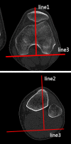
Fig. 1
Fig. 1
Tibial tuberosity–trochlear groove distance (TT–TG) The line 1 was drawn through the bottom of the trochlear groove, while the line 2 was drawn through the middle point of the tibial tubercle. Line 1 and line 2 were perpendicular to the tangent line of the dorsal femoral condylar line (line 3). The TT–TG distance was the distance between the two parallel lines
Fig. 2
Fig. 2
The congruence angle (CA) is the angle with the line drawn through the lower pole of the patella and the deepest point of the chute (line 4) to the line on the side of the bisector defines the tackle angle (line 5)
Fig. 3
Fig. 3
The patellar tilt angle (PTA) is the angle between the extension line of the maximum transverse diameter of the patella (line 6) and the tangent to the posterior condyles (line 7)
Fig. 4
Fig. 4
The medial–lateral width (MLW) is the length between the medial (a) and lateral edge (b) of the epicondyle
Fig. 5
Fig. 5
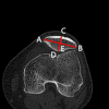
The patellar width (PW) is the length between the medial (A) and lateral edge (B) of the patella. The patellar thickness (PT) is the length between the patellar front polar (C) and back polar (D). The modified Wiberg index is defined as the ratio of the transverse length of the lateral patellar facet (AE) to the medial patellar facet (BE)
Fig. 6
Fig. 6
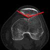
The Wiberg angle is the angle formed by the medial and the lateral patellar facet tangent
Fig. 7
Fig. 7
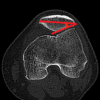
The lateral patellar facet angle is the angle formed by the patellar transverse axis and the lateral patellar facet tangent
Fig. 8
Fig. 8
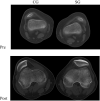
CT scans of patients surgically treated in the study group and conservatively treated in the control group. The scans show remodeling of the patella at the last follow-up in SG patients, which was not seen in CG. CG, control group; SG, study group; Pre, preoperatively; Post, postoperatively

Similar articles

Cited by

References

    1. Sanders TL, Pareek A, Hewett TE, Stuart MJ, Dahm DL, Krych AJ. Incidence of first-time lateral patellar dislocation: a 21-year population-based study. Sports Health. 2018;10(2):146–151. doi: 10.1177/1941738117725055. - DOI - PMC - PubMed
    1. Lewallen LW, McIntosh AL, Dahm DL. Predictors of recurrent instability after acute patellofemoral dislocation in pediatric and adolescent patients. Am J Sports Med. 2013;41(3):575–581. doi: 10.1177/0363546512472873. - DOI - PubMed
    1. Lewallen L, McIntosh A, Dahm D. First-time patellofemoral dislocation: risk factors for recurrent instability. J Knee Surg. 2015;28(4):303–309. doi: 10.1055/s-0034-1398373. - DOI - PubMed
    1. Jaquith BP, Parikh SN. Predictors of recurrent patellar instability in children and adolescents after first-time dislocation. J Pediatr Orthop. 2017;37:484–490. doi: 10.1097/BPO.0000000000000674. - DOI - PubMed
    1. Parikh SN, Nathan ST, Wall EJ, Eismann EA. Complications of medial patellofemoral ligament reconstruction in young patients. Am J Sports Med. 2013;41(5):1030–1038. doi: 10.1177/0363546513482085. - DOI - PubMed

LinkOut - more resources