. 2021 Oct 16;40(1):325.
doi: 10.1186/s13046-021-02137-9.

M6A associated TSUC7 inhibition contributed to Erlotinib resistance in lung adenocarcinoma through a notch signaling activation dependent way

Affiliations

M6A associated TSUC7 inhibition contributed to Erlotinib resistance in lung adenocarcinoma through a notch signaling activation dependent way

Kai Li et al. J Exp Clin Cancer Res. .

Erratum in

Abstract

Background: The small tyrosine kinase inhibitors (TKIs) subversively altered the lung cancer treatments, but patients will inevitably face the therapy resistance and disease recurrence. We aim to explore the potential roles of non-coding RNAs in sensitizing the TKIs effects.

Methods: Multiple cellular and molecular detections were applied to confirm the mechanistic regulations and intracellular connections.

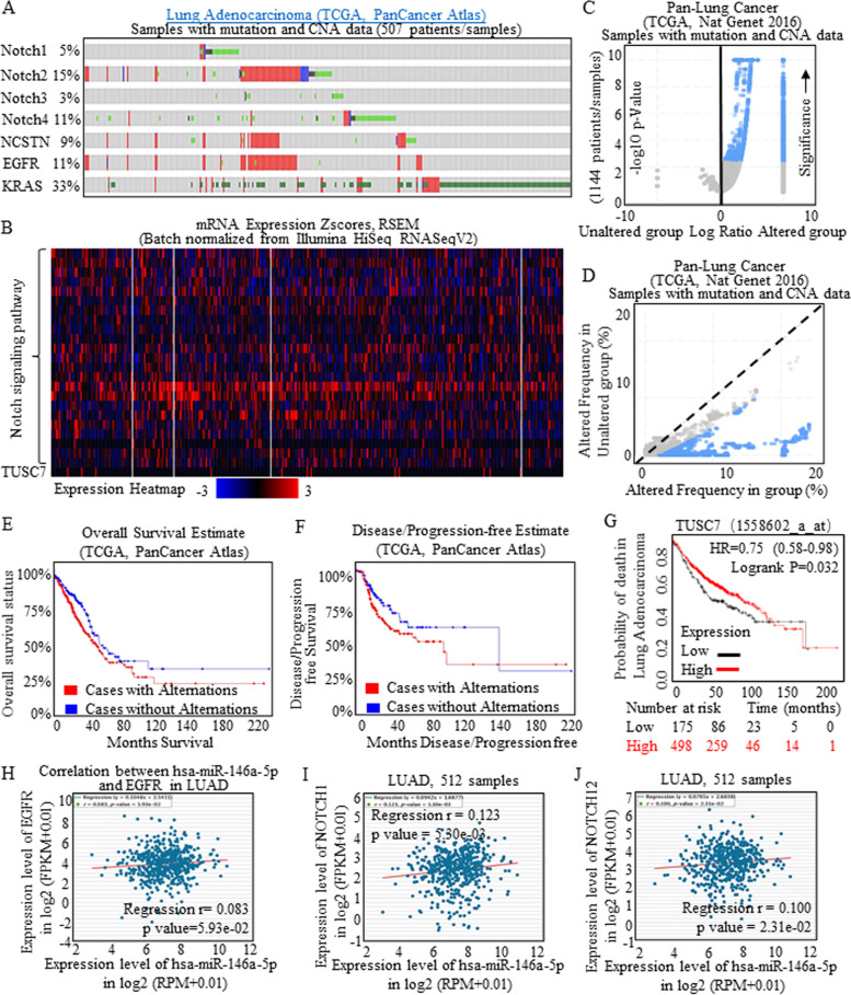
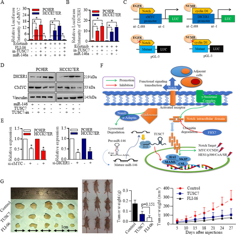
Results: We explored the specific gene features of candidates in association with resistance, and found that m6A controlled the stemness of EMT features through METTL3 and YTHDF2. The miR-146a/Notch signaling was sustained highly activated in a m6A dependent manner, and the m6A regulator of YTHDF2 suppressed TUSC7, both of which contributed to the resistant features. Functionally, the sponge type of TUSC7 regulation of miR-146a inhibited Notch signaling functions, and affected the cancer progression and stem cells' renewal in Erlotinib resistant PC9 cells (PC9ER) and Erlotinib resistant HCC827 cells (HCC827ER) cells. The Notch signaling functions manipulated the cMYC and DICER inner cytoplasm, and the absence of either cMYC or DICER1 lead to TUSC7 and miR-146a decreasing respectively, formed the closed circle to maintain the balance.

Conclusion: PC9ER and HCC827ER cells harbored much more stem-like cells, and the resistance could be reversed by Notch signaling inactivation. The intrinsic miR-146 and TUSC7 levels are monitored by m6A effectors, the alternation of either miR-146 or TUSC7 expression could lead to the circling loop to sustain the new homeostasis. Further in clinics, the combined delivery of TKIs and Notch specific inhibitory non-coding RNAs will pave the way for yielding the susceptibility to targeted therapy in lung cancer.

Keywords: Cancer stem cells; N6-methyladenosine; Notch signaling; Therapy resistance; Tyrosine kinase inhibitors.


Conflict of interest statement

The authors declare that they have no competing interests” in this section.

Figures

Fig. 1
Fig. 1
Aberrant Notch signaling activation in Lung adenocarcinoma. Notch signaling of NOTCH1, NOTCH2, NOTCH3, NOTCH4, DVL1, NUMB, NCSTN, APH1A, SNW1, DTX2, DTX3, DTX1, NCOR2, CTBP2, CTBP1, HDAC1, HDAC2, CIR1, RBPJ, RBPJL, CREBBP, KRAS, MAPK1, MAPK1 were applied for expression level detection, and a total of 507 lung adenocarcinoma samples with mutation and CNA data (TCGA, Pan-Cancer Atlas) were collected. A Expression plots showed the key members of Notch signaling were amplified, together with EGFR overexpression, and KRAS mutations. B Heat map results revealed the universal overexpression of Notch signaling participants, and the TUSC7 result was limited due to the mRNA expression screening system restrictions. Specifically, expression level of TUSC7 showed the reverse consistency with the Notch signaling activation patterns. The TCGA samples data with mutation and CAN from Pan-Lung Cancer (Nat Genet 2016) indicated the grouped enrichment of the Notch signaling factors in Volcano Plots (C), and the changing frequency of each member was consistent with groups alternations (D). E-F The Overall survival and Disease/Progression-free estimates with using Kaplan-Meier analysis showed that Notch signaling activation decreased the survival time, and patients tended to bear relapse or resistance in shorter follow-up periods. G The relative higher level of TUSC7 indicated longer survival time of all lung carcinoma patients significantly, comparting to the lower expressed groups. Sourced data of Star-Base (v3.0 Project) were collected and analyzed, and the results were calculated with using RPM/Log manner, and a total of 512 lung adenocarcinoma samples were enrolled and applied for analysis. H The positive relationship between expression level of miR-146a and level of EGFR was found in total of 512 lung adenocarcinoma samples. Both Notch 1 (I) and Notch 2 (J) were positively correlated to miR-146a expression, pointing to the oncogenic functions of miR-146a
Fig. 2
Fig. 2
Notch inhibition decreased the self-renewal ability of Erlotinib resistant cells and re-sensitized the resistant cells to Erlotinib. A The addition of Erlotinib decreased the ALDH1A1 positive cells of PC9 and HCC827 cells significantly, but did not affect the ratios of Erlotinib resistant PC9ER and HCC827ER cells. B The addition of Erlotinib decreased the spheres number of PC9 and HCC827 cells significantly, but did not affect the number of Erlotinib resistant PC9ER and HCC827ER cells. C Representative images of ALDEFLUOR isolation were detailed exhibited. Two kinds of Notch signaling inhibitors, FLI-06 (inhibitor-1), and γ-Secretase inhibitor (inhibitor-2) were used. 200 nM of inhibitor-1 (D) decreased the self-renewal ability of multiple kinds of lung cancer cells, and 50 nM of inhibitor-2 (E) decreased the self-renewal ability of multiple kinds of lung cancer cells. F Notch signaling inhibition decreased the stem cells’ ratio of the Erlotinib resistant cells significantly, and further, the much-lowered concentration of Notch signaling inhibitor-1, the 20 nM of FLI-06 sensitized both PC9ER and HCC827ER cells to Erlotinib treatment greatly. Erlotinib alone inhibited the Notch signaling slightly, and lowered concentration of FLI-06 mildly inhibited the Notch signaling, but effectively enhanced the Erlotinib functions in PC9ER (Fig. 3G) and HCC827ER cells (Fig. 3H). Combined TUSC7 and Erlotinib decreased the stem cells ratio greatly in both PC9ER and HCC827ER cells (Fig. 3I-J). K-L The stem cells’ renewal suppression evaluation did not show significant differences between TUSC7 alone and the combination of TUSC7 and FLI-06
Fig. 3
Fig. 3
MiR-146a conducted NUMB degradation was blocked by TUSC7 in a sponge combination manner. A The predicated connection sites between TUSC7 and its binding partners showed that miR-146a shared the common untranslated regions. B Recombined miR-146a mimics decreased the Luc-activity of TUSC7 in 293 T cells. MiR-146a decreased the Luc-activity of TUSC7 in PC9ER cells (C) and HCC827ER cells (D). E Blotting results referring to RNA pull-down test showed the connection between TUSC7 and NUMB in PC9ER and HCC827ER cells. F RNA immunoprecipitation revealed that TUSC7 was enriched with NUMB expression in PC9ER cells (Left) and HCC827ER cells (Right), and the IgG was set as the immunoprecipitation control, the MALAT1 was set as the primer control. G Informatic screening of the potential miRNAs’ targets suggested that miR-146a may bind to NUMB. H The expression level of NUMB with 526 cancer and 59 normal samples in LUAD, and the expressions with 501 cancer and 49 normal samples in LUSC were evaluated, and NUMB decreased greatly in cancer group. I The co-transfection of miR-146a mimics and the wide-type reporter plasmid strongly reduced the EGFP intensity, and miR-146a mimics reduced nearly 40% of the TUSC7 luciferase intensity, but not happened in mutant-type reporter plasmid. J TUSC7 alone did not reduce the EGFP activity of NUMB. K MiR-146a decreased the NUMB expression, which could be rescued by TUSC7, and the TUSC7 inhibition (TUSC7-in) also decreased the NUMB level
Fig. 4
Fig. 4
m6A status was associated with TUSC7 inhibition and snail relating miR-146a overexpression. A M6A levels of RNAs from resistant cells were statistically more abundant than sensitive original cells. METTL3 affected the miR-146a level (B), and YTHDF2 affected TUSC7 level (C). D-E The results were all confirmed by using the lentiviral based METTL3/YTHDF2 knock-down systems. F-G Dysregulated METTL3 and YTHDF2 affected the m6A, and then determined different EMT and stemness feature in resistant PC9ER cells and HCC827ER cells. H-I METTL3 inhibition decreased m6A at Snai1. J Snai1 inhibition failed to activate the miR-146a promoter activity. K The m6A at TUSC7 level increased in resistant cells, and the recognition of TUSC7 m6A peak by YTHDF2 degraded and downregulated the TUSC7 expression. L The Me-RIP assay confirmed that the high abundance of m6A modification in cells with YTHDF2 inhibition
Fig. 5
Fig. 5
TUSC7 sensitized Erlotinib treatment and formed feedback loop with miR-146/Notch signaling cascade. A Notch signaling inhibition resulted in cMYC promoter activity decreasing, and TUSC7 inhibition strongly increased the activity. B Inhibition of TUSC7 increased miR-146a expression, and then stimulated the DICER1 activity. C The scheme image represented the cMYC promoter detection modes. D Western blotting confirmed that in PC9ER and HCC827ER cells, miR-146 and TUSC7 acted the opposite way to promote the DICER1/CCND1 expression. E TUSC7 and miR-146a was sustained by cMYC (Left) and DICER1 (Right) respectively. F The regulative signaling pathways were drafted and illustrated for detailed exhibition. G In vivo study confirmed the effective inhibition of TUSC7 exhibited on tumor growth, and the Notch signaling inactivation by using FLI-06 also suppressed the in vivo tumor expansion

References

    1. Bray F, Ferlay J, Soerjomataram I, Siegel RL, Torre LA, Jemal A. Global cancer statistics 2018: GLOBOCAN estimates of incidence and mortality worldwide for 36 cancers in 185 countries. CA Cancer J Clin. 2018;68(6):394–424. doi: 10.3322/caac.21492. - DOI - PubMed
    1. Bogos K, Kiss Z, Gálffy G, Tamási L, Ostoros G, Müller V, et al. Revising Incidence and Mortality of Lung Cancer in Central Europe: An Epidemiology Review From Hungary. Front Oncol. 2019;9:1051. - PMC - PubMed
    1. Zheng R, Zeng H, Zuo T, Zhang S, Qiao Y, Zhou Q, et al. Lung cancer incidence and mortality in China, 2011. Thorac Cancer. 2016;7(1):94–99. doi: 10.1111/1759-7714.12286. - DOI - PMC - PubMed
    1. Chen WQ, Zuo TT, Zheng RS, Zeng HM, Zhang SW, He J. Lung cancer incidence and mortality in China in 2013. Zhonghua Zhong Liu Za Zhi. 2017;39(10):795–800. - PubMed
    1. Raaschou-Nielsen O, Andersen ZJ, Beelen R, Samoli E, Stafoggia M, Weinmayr G, et al. Air pollution and lung cancer incidence in 17 European cohorts: prospective analyses from the European study of cohorts for air pollution effects (ESCAPE) Lancet Oncol. 2013;14(9):813–822. doi: 10.1016/S1470-2045(13)70279-1. - DOI - PubMed