Review
. 2021 Nov:47:102164.
doi: 10.1016/j.redox.2021.102164. Epub 2021 Oct 12.

Mitochondrial iron-sulfur clusters: Structure, function, and an emerging role in vascular biology

Affiliations
Review

Mitochondrial iron-sulfur clusters: Structure, function, and an emerging role in vascular biology

Austin D Read et al. Redox Biol. 2021 Nov.

Abstract

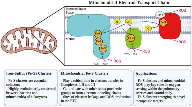
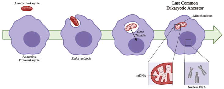
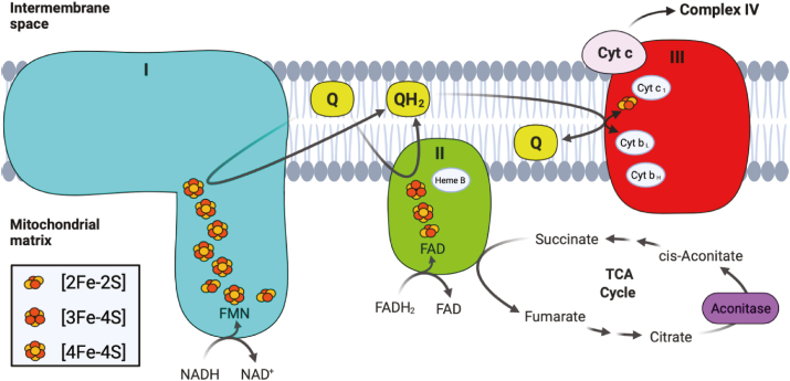
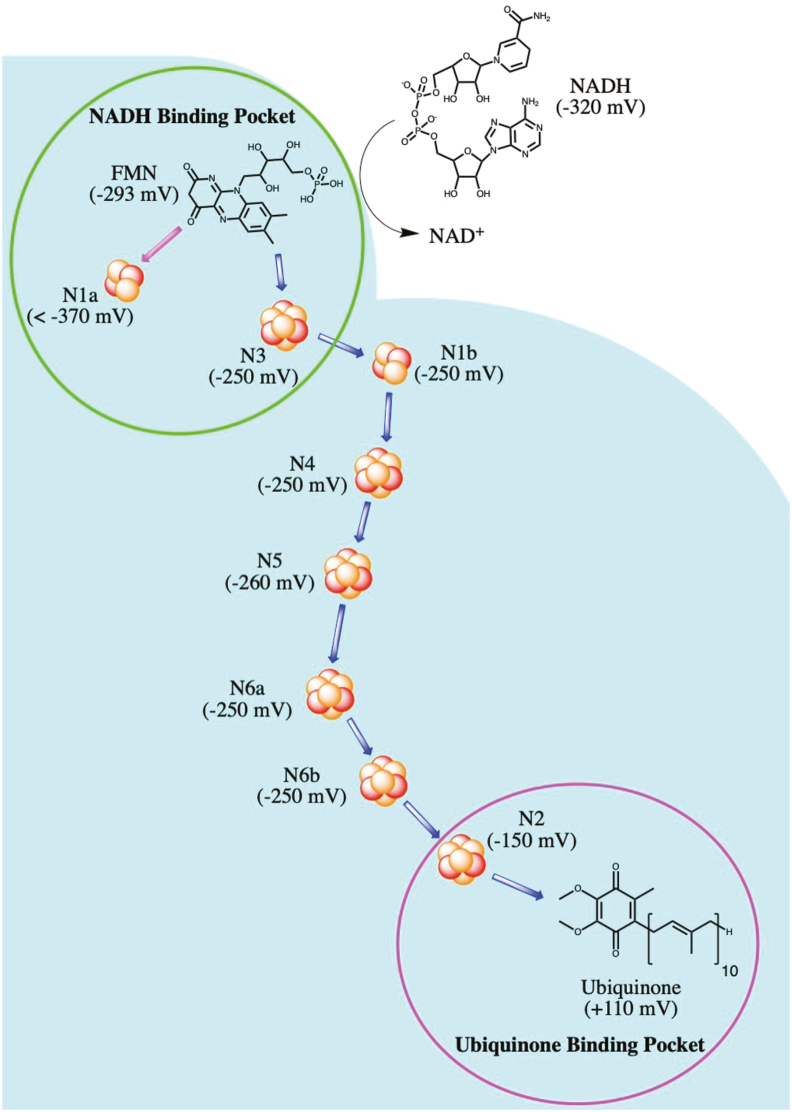
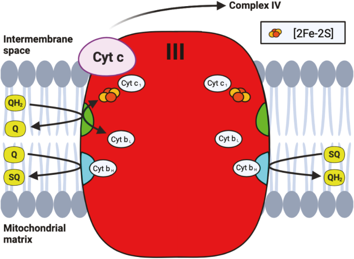
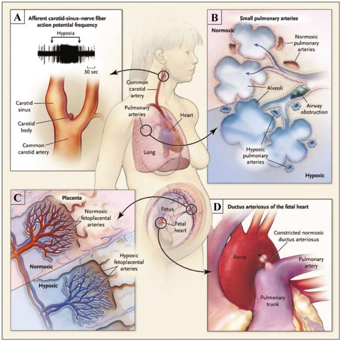
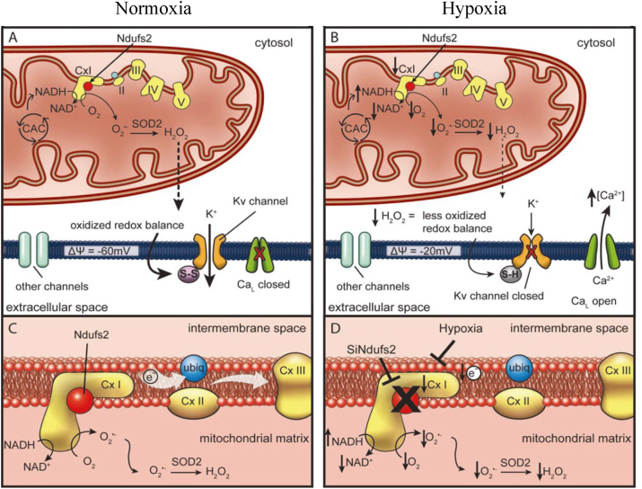
Iron-sulfur (Fe-S) clusters are essential cofactors most commonly known for their role mediating electron transfer within the mitochondrial respiratory chain. The Fe-S cluster pathways that function within the respiratory complexes are highly conserved between bacteria and the mitochondria of eukaryotic cells. Within the electron transport chain, Fe-S clusters play a critical role in transporting electrons through Complexes I, II and III to cytochrome c, before subsequent transfer to molecular oxygen. Fe-S clusters are also among the binding sites of classical mitochondrial inhibitors, such as rotenone, and play an important role in the production of mitochondrial reactive oxygen species (ROS). Mitochondrial Fe-S clusters also play a critical role in the pathogenesis of disease. High levels of ROS produced at these sites can cause cell injury or death, however, when produced at low levels can serve as signaling molecules. For example, Ndufs2, a Complex I subunit containing an Fe-S center, N2, has recently been identified as a redox-sensitive oxygen sensor, mediating homeostatic oxygen-sensing in the pulmonary vasculature and carotid body. Fe-S clusters are emerging as transcriptionally-regulated mediators in disease and play a crucial role in normal physiology, offering potential new therapeutic targets for diseases including malaria, diabetes, and cancer.

Keywords: Drug target; Electron transport chain; Epigenetics; Fe-S cluster; Mitochondria; Oxygen-sensing.


Conflict of interest statement

The authors declare that they have no known competing financial interests or personal relationships that could have appeared to influence the work reported in this paper.

Figures

Image 1
Graphical abstract
Fig. 1
Fig. 1
The most common forms of Fe–S clusters found in biological systems: [2Fe–2S], [3Fe–4S] and [4Fe–4S]. In all structures shown above, the iron and sulfur atoms that are a part of the cluster are shown in red and ligating amino acid residues are shown in black. (For interpretation of the references to colour in this figure legend, the reader is referred to the Web version of this article.)
Fig. 2
Fig. 2
Overview of endosymbiont hypothesis. This universally accepted theory of mitochondrial origins posits that the anaerobic proto-eukaryote engulfed the aerobic prokaryote, most closely related to the order Rickettsiales. There was sizeable transfer of genes encoding mitochondrial proteins to the nuclear genome and elimination of redundancy. with current mitochondrial genomes encoding as few as 5 genes. The acquisition of the mitochondria resulted in the last common eukaryotic ancestor, from which all current eukaryotes evolved.
Fig. 3
Fig. 3
Aconitase reaction mechanism. The isomerization reaction of citrate (shown in blue) to cis-Aconitate (magenta) is facilitated by the [4Fe–4S] cluster found within aconitase (shown in red). The addition of another labile iron (Feα) transforms inactive aconitase into its active form, and coordinates with water molecules and oxygen atoms of citrate to facilitate enzyme catalysis. (For interpretation of the references to colour in this figure legend, the reader is referred to the Web version of this article.)
Fig. 4
Fig. 4
Simplified version of the mitochondrial ETC, showing Complexes I (blue), II (green), and III (red). The electron donors NADH and FADH2 are shown reducing Complexes I and respectively, as well as the electron carriers' ubiquinone (Q) and ubiquinol (QH2). Besides the Fe–S clusters, other redox prosthetic groups within each complex are also shown, including FMN and FAD, as well as the heme moieties a part of the cytochrome proteins. Intermediates within the TCA cycle that interact with Complex II and aconitase (purple) are also shown. (For interpretation of the references to colour in this figure legend, the reader is referred to the Web version of this article.)
Fig. 5
Fig. 5
Fe–S chain within Complex I. The midpoint redox potential at pH of 7 (as found in B. taurus) is listed for each cluster, as well as NADH, FMN, and ubiquinone. The NADH and ubiquinone binding sites are depicted in green and magenta circles, respectively, and the main route of electron transfer is depicted by blue arrows. (For interpretation of the references to colour in this figure legend, the reader is referred to the Web version of this article.)
Fig. 6
Fig. 6
Q-cycle within Complex III. Ubiquinol (QH2) is oxidized at the Qo site (shown in green), passing electrons to either cytochrome c (cyt c) through the [2Fe–2S] cluster within the ISP and cytochrome cl (cyt c1), or to the Qisite (shown in blue) through cyt bL and bH. As shown on the left side of the complex, ubiquinone (Q) is reduced to ubisemiquinone (SQ) at the Qi site after a single round of the Q-cycle, while a second round of the cycle reduces SQ to QH2, as shown on the right side of the complex. (For interpretation of the references to colour in this figure legend, the reader is referred to the Web version of this article.)
Fig. 7
Fig. 7
The specialized tissues of the Homoeostatic Oxygen Sensing System (HOSS). A. The carotid body, (located at the bifurcation of the carotid artery) increases the frequency of action potentials upon exposure to hypoxia. B. Pulmonary arteries constrict in hypoxia to divert blood to regions of the lung with better oxygenation (ventilation-perfusion matching). C. Hypoxic Fetoplacental vasoconstriction optimizes maternal and fetal perfusion matching. D. The ductus arteriosus rapidly constricts in response to nomioxia postnatally, properly separating the pulmonary and systemic circulations. Adapted with permission from [138] Weir et al. (2005) NEJM.
Fig. 8
Fig. 8
PASMC mitochondria during normoxia and hypoxia. A. Under normoxic conditions, oxygen radicals produced at ETC Complex I are converted to H2O2 by SOD2, with this ROS production leading to oxidation of sulfhydryl groups in the Kv channel, forming disulfide bridges (S–S) and increasing the channel's open-state probability. B. With a switch to hypoxia, production of superoxide is decreased due to decreased uncoupled electron transport. The resultant decrease in H2O2 and the accumulation of NADH depolarize the cell, closing the Kv channels, with subsequent opening of calcium channels causing vasoconstriction. C. Optimal function of Complex I requires intact Ndufs2. D. Inhibition of Complex I, via hypoxia or siRNA targeting Ndufs2, results in a more reduced redox state. Adapted with permission from [173] Dunham-Snary et al. (2019) Circ. Res.

References

    1. Burén S., Jiménez-Vicente E., Echavarri-Erasun C., Rubio L.M. Biosynthesis of nitrogenase cofactors. Chem. Rev. 2020;120(12):4921–4968. - PMC - PubMed
    1. Luo Y., Ergenekan C.E., Fischer J.T., Tan M.L., Ichiye T. The molecular determinants of the increased reduction potential of the rubredoxin domain of rubrerythrin relative to rubredoxin. Biophys. J. 2010;98(4):560–568. - PMC - PubMed
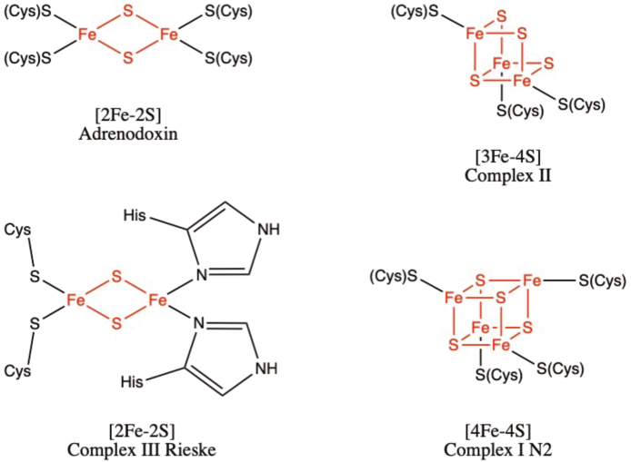
    1. Ewen K.M., Ringle M., Bernhardt R. Adrenodoxin–a versatile ferredoxin. IUBMB Life. 2012;64(6):506–512. - PubMed
    1. Ali M.E., Nair N.N., Retegan M., Neese F., Staemmler V., Marx D. The iron–sulfur core in Rieske proteins is not symmetric. JBIC Journal of Biological Inorganic Chemistry. 2014;19(8):1287–1293. - PubMed
    1. Ohnishi T. Iron–sulfur clusters/semiquinones in Complex I, Biochimica et Biophysica Acta (BBA) Bioenergetics. 1998;1364(2):186–206. - PubMed

Publication types