. 2022 Feb;22(2):344-370.
doi: 10.1111/ajt.16868. Epub 2021 Nov 1.

Preclinical models versus clinical renal ischemia reperfusion injury: A systematic review based on metabolic signatures

Affiliations

Preclinical models versus clinical renal ischemia reperfusion injury: A systematic review based on metabolic signatures

Lente J S Lerink et al. Am J Transplant. 2022 Feb.

Abstract

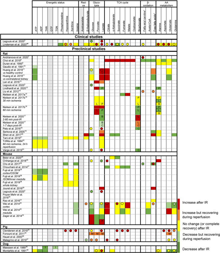
Despite decennia of research and numerous successful interventions in the preclinical setting, renal ischemia reperfusion (IR) injury remains a major problem in clinical practice, pointing toward a translational gap. Recently, two clinical studies on renal IR injury (manifested either as acute kidney injury or as delayed graft function) identified metabolic derailment as a key driver of renal IR injury. It was reasoned that these unambiguous metabolic findings enable direct alignment of clinical with preclinical data, thereby providing the opportunity to elaborate potential translational hurdles between preclinical research and the clinical context. A systematic review of studies that reported metabolic data in the context of renal IR was performed according to the PRISMA guidelines. The search (December 2020) identified 35 heterogeneous preclinical studies. The applied methodologies were compared, and metabolic outcomes were semi-quantified and aligned with the clinical data. This review identifies profound methodological challenges, such as the definition of IR injury, the follow-up time, and sampling techniques, as well as shortcomings in the reported metabolic information. In light of these findings, recommendations are provided in order to improve the translatability of preclinical models of renal IR injury.

Keywords: animal models; delayed graft function; ischemia reperfusion injury; kidney failure / injury; kidney transplantation / nephrology; metabolomics; translational research / science.


Figures

FIGURE 1
FIGURE 1
Illustration of the contrasting postreperfusion metabolic responses of kidney donor grafts with delayed graft function (DGF, IR injury, red curve) and grafts recovering without IR injury (no DGF) (green curve: living donor graft [intermediate ischemic period], blue curve: deceased donor graft [prolonged ischemic period]). The dashed line reflects the normal, non‐ischemic kidney. Figures adapted from Lindeman et al. Curves represent renal vein levels of lactate (glycolysis), uracil (cellular damage), hypoxanthine (ATP catabolism and metabolic incompetence), and isovalerylcarnitine (intermediate of branched‐chain amino acid oxidation [autophagia]). *Decrease at end of measurement window may reflect depletion of ATP pool
FIGURE 2
FIGURE 2
Relative differences in renal tissue (boxes) and blood (spheres, note: often systemic if no data on kidney‐specific samples) metabolite levels between control kidneys (healthy/intact control, sham‐surgery control, or contralateral kidney: see Table 2) and kidneys after ischemia reperfusion (IR). If no control data were available, data were excluded. Metabolites marked in grey were not reported for tissue or blood samples in any of the preclinical studies. Red = increase after IR. Orange = increase but recovering during reperfusion (requires multiple samples during reperfusion). Yellow = no change (or complete recovery) after IR. Light green = decrease but recovering during reperfusion (requires multiple samples during reperfusion). Green = decrease after IR. TAN: total adenine nucleotides (ATP+ADP+AMP). TGN: total guanine nucleotides (GTP+GDP+GMP). *: variety of fatty acids, results on overall trend. #: additional data on tissue levels are reported in the supplementary data, but no formal statistical evaluation was performed. OSOM: outer stripes of outer medulla. ISOM: inner stripes of outer medulla. †: ischemia time dependent. P: metabolite/pyruvate ratio. L: metabolite/lactate ratio. B: metabolite/bicarbonate ratio. C: in control (sham‐operated animal or contralateral kidney), metabolite levels showed similar dynamics following surgery. M: measured using microdialysis
FIGURE 3
FIGURE 3
Recommendations to improve the translatability of preclinical models of renal ischemia reperfusion (IR) injury. Ischemic injury should be induced unilaterally in order to avoid interference caused by uremia. Appropriate discrimination between IR damage and IR injury critically relies on the organ‐specific assessment of metabolic competence (i.e., prolonged normoxic glycolysis, see outline in Figure 1). Kidney‐specific metabolic profiling of the injured kidney can be achieved through renal vein–specific blood sampling using the spermatic vein as access., Microdialysis is a potential alternative to arteriovenous sampling, but has not yet been validated for this purpose. Ureterostomy allows for selective functional monitoring of the injured kidney. The model critically relies on optimized ischemia times in order to achieve actual IR injury and avoid excess incidences of IR damage or non‐function. It is anticipated that successful implementation of these prerequisites relies on use of rats or larger laboratory animals. Illustration by Manon Zuurmond

References

    1. Lefer DJ, Bolli R. Development of an NIH Consortium for PreclinicAl AssESsment of CARdioprotective Therapies (CAESAR): a paradigm shift in studies of infarct size limitation. J Cardiovasc Pharmacol Ther. 2011;16(3–4):332‐339. doi:10.1177/1074248411414155 - DOI - PubMed
    1. Cavaillé‐Coll M, Bala S, Velidedeoglu E, et al. Summary of FDA workshop on ischemia reperfusion injury in kidney transplantation. Am J Transplant. 2013;13(5):1134‐1148. doi:10.1111/ajt.12210 - DOI - PubMed
    1. Legouis D, Ricksten SE, Faivre A, et al. Altered proximal tubular cell glucose metabolism during acute kidney injury is associated with mortality. Nat Metab. 2020;2(8):732‐743. doi:10.1038/s42255-020-0238-1 - DOI - PubMed
    1. Lindeman JH, Wijermars LG, Kostidis S, et al. Results of an explorative clinical evaluation suggest immediate and persistent post‐reperfusion metabolic paralysis drives kidney ischemia reperfusion injury. Kidney Int. 2020;98(6):1476‐1488. doi:10.1016/j.kint.2020.07.026 - DOI - PubMed
    1. Van Os S, De Abreu R, Hopman J, Wethly K, Liem D, Van De Bor M. Purine and pyrimidine metabolism and electrocortical brain activity during hypoxemia in near‐term lambs. Pediatr Res. 2004;55(6):1018‐1025. doi:10.1203/01.PDR.0000125261.99069.D5 - DOI - PubMed

Publication types