. 2021 Dec;78(6):1902-1913.
doi: 10.1161/HYPERTENSIONAHA.121.18190. Epub 2021 Oct 18.

Enhanced Sp1/YY1 Expression Directs CBS Transcription to Mediate VEGF-Stimulated Pregnancy-Dependent H2S Production in Human Uterine Artery Endothelial Cells

Affiliations

Enhanced Sp1/YY1 Expression Directs CBS Transcription to Mediate VEGF-Stimulated Pregnancy-Dependent H2S Production in Human Uterine Artery Endothelial Cells

Jin Bai et al. Hypertension. 2021 Dec.

Abstract

[Figure: see text].

Keywords: endothelial cells; humans; hydrogen sulfide; pregnancy; uterine artery; vascular endothelial growth factor A.


Conflict of interest statement

Disclosure: The authors declare no conflict of interest.

Figures

Fig. 1:
Fig. 1:. VEGF stimulates pregnancy dependent human uterine artery endothelial CBS expression.
A: Primary human uterine artery endothelial cells (hUAEC) were treated with 10 ng/mL vascular endothelial growth factor (VEGF) for up to 2 days to assess cystathionine β-synthase (CBS) and cystathionine γ-lyase (CSE) mRNA. Bars with different letters differ significantly (p<0.05). Data (means ± SEM) were collected from different hUAEC preparations from different nonpregnnat (NP) and pregnant (P) women. B: P hUAEC were treated with or without actinomycin D (5 μg/ml). Total RNA samples were extracted at 0, 3, 6, 9 and 12 h post actinomycin D. CBS and L19 were analyzed using real time quantitative reverse transcription-PCR (RT-qPCR). Data (means ± SEM, n=3) were presented as a percentage of relative CBS expression at time 0. C: Both non-pregnant (NP) and P hUAEC were transfected with luciferase constructs driven by wild-type human CBS promoter (−753/+161) or human CSE promoter (−942/+98) and co-transfected with the thymidine kinase-Renilla luciferase vector. After VEGF (10 ng/ml) treatment for 24 h, trans-activation of CBS or CSE promoter was measured by luciferase reporter gene expressions as a ratio of firefly/Renilla luciferase activities. Data (mean ± SEM. N=3) were presented as fold of controls. * p<0.05 and ** p<0.01. D: P hUAEC were transfected with luciferase constructs driven by wild-type human CBS promoter or CSE promoter and co-transfected with the thymidine kinase-Renilla luciferase vector, treated with increasing doses (0-100 ng/ml) of VEGF for 24 h. Data were summarized as mean±SEM from three independent experiments; Bars with different letters differ significantly (p < 0.05).
Fig. 2:
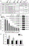
Fig. 2:. Sp1/YY1 in VEGF-stimulated CBS transcription.
A: Putative Sp1 and YY1 binding sites in human CBS promoter and primers designed to amply amplicons containing specific Sp1/YY1 sites for chromatin immunoprecipitation-PCR. P hUAEC were transfected with luciferase reporter constructs driven by wild-type human CBS promoter (−753/+161) or its 5’-deletions (B), and wild type (WT) pCBS(−574).Luc construct or its mutations in the Sp1d, YY1, or both sites (C), and with thymidine kinase-Renilla luciferase vector as internal control. After treatment with VEGF (10 ng/ml) for 24 h, trans-activation of CBS promoter was measured. Data (means ± SEM, n=3) were expressed as fold of baseline WT CBS promoter activity. Bars with different letters differ significantly (p < 0.05). ***, p<0.001 vs controls.
Fig. 3:
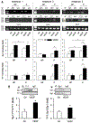
Fig. 3:. Sp1/YY1 interactions with CBS promoter.
A: Binding of Sp1 and YY1 to human CBS promoter. Nonpregnant (NP) and pregnant (P) hUAEC were treated with or without VEGF (10 ng/ml, 24 h). Sheared DNA from the cells were subjected to chromatin immunoprecipitation (ChIP) using Sp1 or YY1 antibodies; IgG was used as negative controls. Sp1 and YY1 binding with specific sites were amplified by PCR using primers as designed in Fig. 2A. Data (means ± SEM) were summarized as fold of control from three experiments using cells from different women. *, p< 0.05 vs control. B. Sp1 and YY1 association. P hUAEC were treated with or without VEGF (10 ng/ml, 30 min). Proteins (1 mg/sample) were used immunoprecipitation (IP) with Sp1 or YY1 antibodies and IgG was used as control. The IP samples were immunoblotted with Sp1 or YY1. Levels of Sp1 bound YY1 and YY1 bound Sp1 were summarized as means ± SEM from NP or P hUAEC from three different women. *, p<0.05 vs control.
Fig. 4:
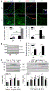
Fig. 4:. Sp1 and YY1 proteins in human uterine artery and effects of VEGF on hUAEC Sp1/YY1 expresison.
A: Paraffin-embedded human uterine artery rings were immunolabled by specific Sp1 or YY1 antibodies and CD31 antibody for labeling endothelial cells. Sections were mounted with DAPI to label nuclei and examined under confocal microscopy. Images were taken to determine Sp1 and YY1 proteins (relative green fluorescence intensity) in endothelium (EC) and smooth muscle (SM) using Image J and summarized in graphs. *, p < 0.05, **, p < 0.01, ***, p < 0.001 vs NP controls. B: Proteins (20 μg/lane) from NP an P hUAEC (n=3/group) were used for immunoblotting with Sp1 and YY1 antibodies and β-actin was measured as loading control. Data were summarized as means ± SEM and presented as fold of NP. *, p<0.05, ***, p<0.01 vs NP. C: P hUAEC were treated with VEGF (10 ng/ml, 3 days) or with increasing concentrations (0–100 ng/ml) of VEGF for 2 days to assess Sp1 and YY1 proteins by immunoblotting. Data were obtained from three experiments using cells from different women and calculated as means ± SEM (fold of baseline). Small and capital letter on bars represent differences in Sp1 and YY1, respectively, and bars with different letter differ significantly (p <0.05).
Fig. 5:
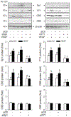
Fig. 5:. Sp1/YY1 knockdown on VEGF-stimulated CBS/CSE expression.
P hUAEC were infected with or without lentiviruses (10 MOI) of Sp1 (siSp1) or YY1 (siYY1) siRNAs for 72 h; lentivirus of scramble siRNA (siCtl) as control. The cells were then treated with or without VEGF (10 ng/ml) for 48 h. Cell lysates (20 μg/lane) were immunoblotted for Sp1, YY1, CBS, and CSE; β-actin was measured as loading control. Data were obtained from three experiments using cells from different women and calculated as means ± SEM (fold of baseline). Bars with different letters differ significantly (p< 0.05).
Fig. 6:
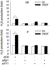
Fig. 6:. Sp1/YY1 knockdown on VEGF-stimulated H2S production.
NP and P hUAEC were infected with or without lentiviruses (10 MOI) of Sp1 (siSp1) or YY1 (siYY1) siRNAs for 72 h; lentivirus of scramble siRNA (siCtl) as control. The cells were then treated with or without VEGF (10 ng/ml) for 48 h. Protein extracts (1 × 10⁶ cells) were used to determine hydrogen sulfide (H2S) production by methylene blue assay. Data (means ± SEM) were collected from NP or P hUAEC (n=3/group) from different women. ***, p < 0.001 vs control.

Similar articles

Cited by

References

    1. Sanghavi M, Rutherford JD. Cardiovascular physiology of pregnancy. Circulation. 2014;130:1003–1008. doi: 10.1161/CIRCULATIONAHA.114.0090292. - DOI - PubMed
    1. Thaler I, Manor D, Itskovitz J, Rottem S, Levit N, Timor-Tritsch I, Brandes JM. Changes in uterine blood flow during human pregnancy. Am J Obstet Gynecol. 1990;162:121–5. doi: 10.1016/0002-9378(90)90834-t. - DOI - PubMed
    1. Gluckman PD, Hanson MA, Cooper C, Thornburg KL. Effect of in utero and early-life conditions on adult health and disease. N Engl J Med. 2008;359:61–73. doi: 10.1056/NEJMra0708473 - DOI - PMC - PubMed
    1. Magness RR, Shaw CE, Phernetton TM, Zheng J, Bird IM. Endothelial vasodilator production by uterine and systemic arteries. Ii. Pregnancy effects on NO synthase expression. Am J Physiol. 1997;272:H1730–1740. doi: 10.1152/ajpheart.1997.272.4.H1730 - DOI - PubMed
    1. Nelson SH, Steinsland OS, Suresh MS, Lee NM. Pregnancy augments nitric oxide-dependent dilator response to acetylcholine in the human uterine artery. Hum Reprod. 1998;13:1361–1367 - PubMed

Publication types