. 2022 Jun;18(6):1350-1366.
doi: 10.1080/15548627.2021.1976965. Epub 2021 Oct 17.

Mechanistic insights into an atypical interaction between ATG8 and SH3P2 in Arabidopsis thaliana

Affiliations

Mechanistic insights into an atypical interaction between ATG8 and SH3P2 in Arabidopsis thaliana

Shuangli Sun et al. Autophagy. 2022 Jun.

Abstract

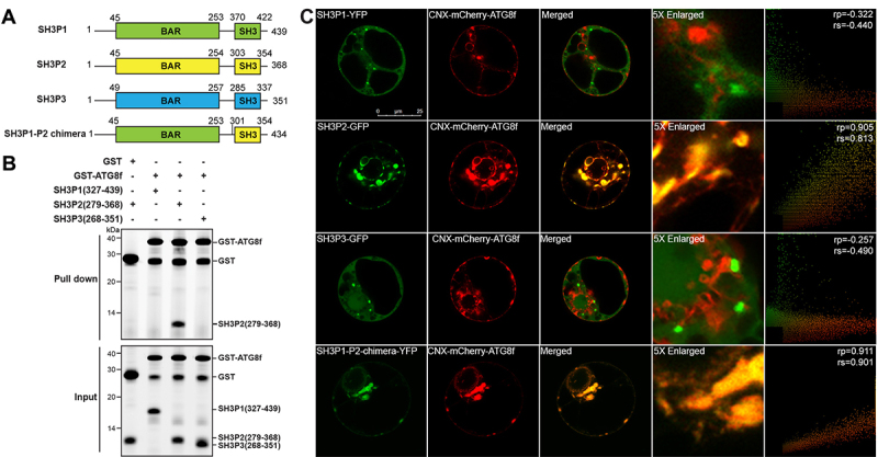
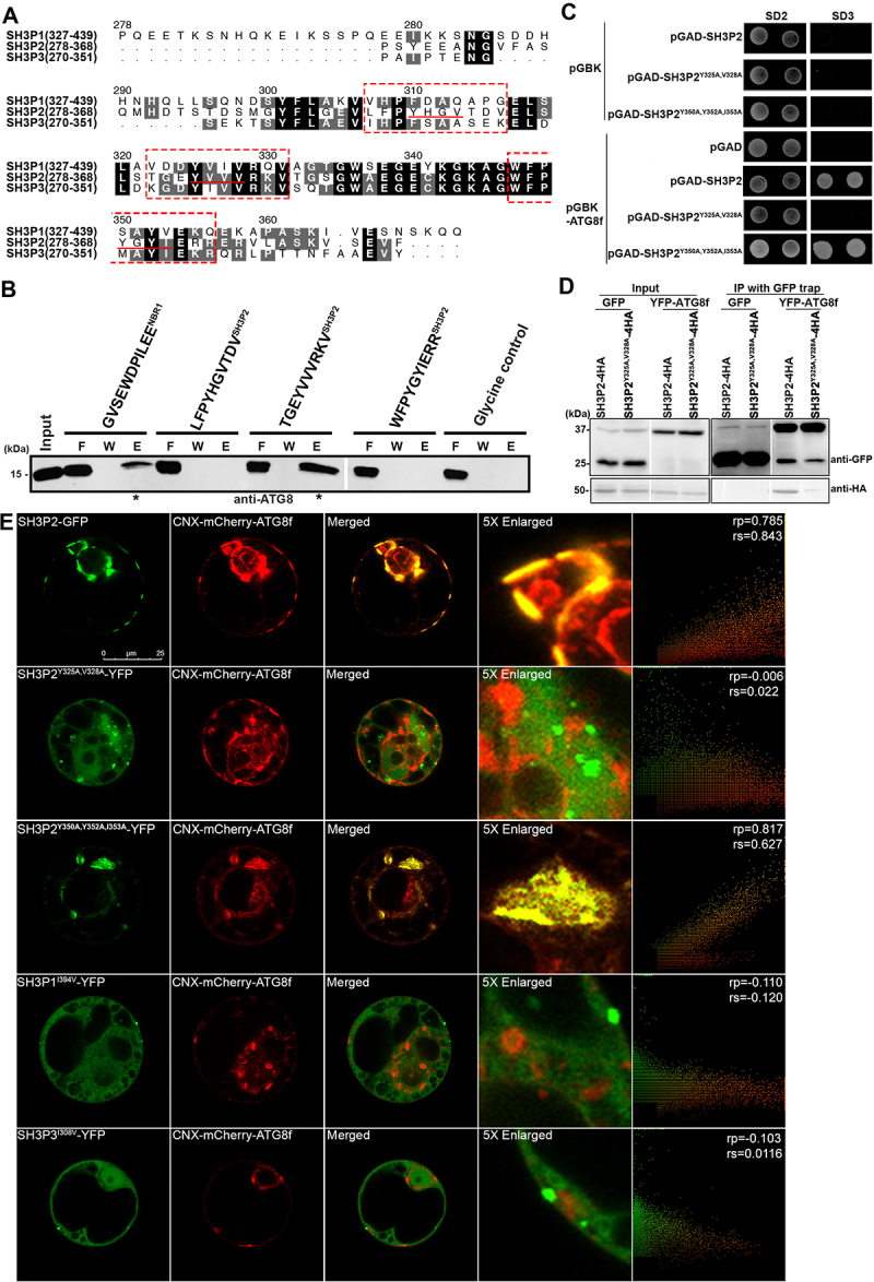
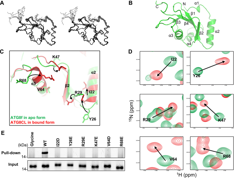
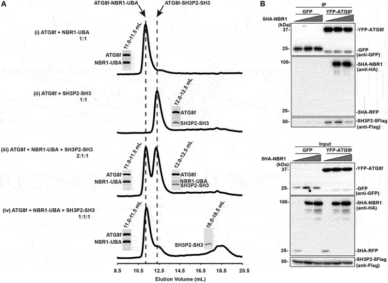
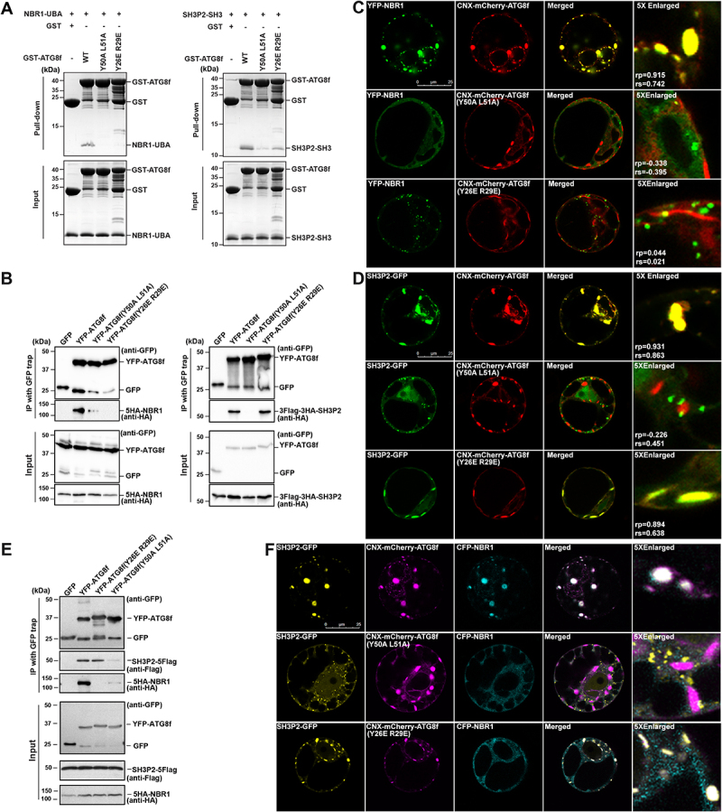
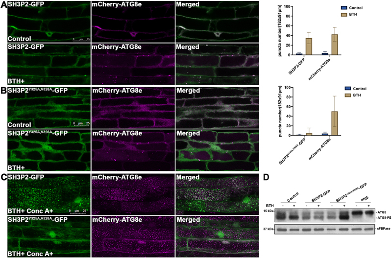
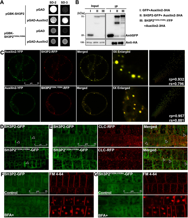
In selective macroautophagy/autophagy, cargo receptors are recruited to the forming autophagosome by interacting with Atg8 (autophagy-related 8)-family proteins and facilitate the selective sequestration of specific cargoes for autophagic degradation. In addition, Atg8 interacts with a number of adaptors essential for autophagosome biogenesis, including ATG and non-ATG proteins. The majority of these adaptors and receptors are characterized by an Atg8-family interacting motif (AIM) for binding to Atg8. However, the molecular basis for the interaction mode between ATG8 and regulators or cargo receptors in plants remains largely unclear. In this study, we unveiled an atypical interaction mode for Arabidopsis ATG8f with a plant unique adaptor protein, SH3P2 (SH3 domain-containing protein 2), but not with the other two SH3 proteins. By structure analysis of the unbound form of ATG8f, we identified the unique conformational changes in ATG8f upon binding to the AIM sequence of a plant known autophagic receptor, NBR1. To compare the binding affinity of SH3P2-ATG8f with that of ATG8f-NBR1, we performed a gel filtration assay to show that ubiquitin-associated domain of NBR1 outcompetes the SH3 domain of SH3P2 for ATG8f interaction. Biochemical and cellular analysis revealed that distinct interfaces were employed by ATG8f to interact with NBR1 and SH3P2. Further subcellular analysis showed that the AIM-like motif of SH3P2 is essential for its recruitment to the phagophore membrane but is dispensable for its trafficking in endocytosis. Taken together, our study provides an insightful structural basis for the ATG8 binding specificity toward a plant-specific autophagic adaptor and a conserved autophagic receptor.Abbreviations: ATG, autophagy-related; AIM, Atg8-family interacting motif; BAR, Bin-Amphiphysin-Rvs; BFA, brefeldin A; BTH, benzo-(1,2,3)-thiadiazole-7-carbothioic acid S-methyl ester; CCV, clathrin-coated-vesicle; CLC2, clathrin light chain 2; Conc A, concanamycin A; ER, endoplasmic reticulum; LDS, LIR docking site; MAP1LC3/LC3, microtubule associated protein 1 light chain 3; LIR, LC3-interacting region; PE, phosphatidylethanolamine; SH3P2, SH3 domain containing protein 2; SH3, Src-Homology-3; UBA, ubiquitin-associated; UIM, ubiquitin-interacting motif.

Keywords: ATG8 interacting motif; Arabidopsis ATG8; NBR1; SH3P2; selective autophagy.


Figures

Figure 1.
Figure 1.
SH3P2, but not SH3P1 and SH3P3, binds to Arabidopsis ATG8f. (A) Domain organization of Arabidopsis SH3Ps proteins. SH3P1, SH3P2 and SH3P3 all contain an N-terminal BAR domain and a C-terminal SH3 domain. (B) GST affinity-isolation assay. 1 µM SH3 domain protein of SH3P1, SH3P2 and SH3P3 was mixed with 1 µM GST-ATG8f and incubated with glutathione resin (upper panel) respectively. The bound protein was eluted with 10 mM glutathione, and then pre-stained by Instant-Bands and analyzed by SDS-PAGE (lower panel). Our results suggested that GST-ATG8f only interacted with the SH3 domain of SH3P2 but not with the SH3 domains of SH3P1 and SH3P3. (C) SH3Ps-GFP/YFP, or SH3P1-P2-chimera-YFP, and CNX-mCherry-ATG8f were transiently co-expressed in Arabidopsis protoplasts. Confocal analysis showed that SH3P2, but not SH3P1 and SH3P3, colocalized with CNX-mCherry-ATG8f. Similar results were obtained from three different independent experiments. The right column shows the scatterplot images obtained from ImageJ with the PSC colocalization plug-in. The linear Pearson correlation coefficient (rp) and the nonlinear Spearman correlation coefficient (rs) indicate the extent of colocalization with the value of +1.0 for complete colocalization.
Figure 2.
Figure 2.
SH3P2 contains an AIM-like motif for interaction with ATG8f. (A)The sequences of the SH3 domain of SH3Ps protein in Arabidopsis were aligned by the Clustal-Omega algorithm [92] implemented in the program SEAVIEW [93]. Three AIM-like sequences (underlined) were identified in SH3P2. Residues were numbered according to the Arabidopsis SH3P2 sequence. (B) The purified ATG8f protein was incubated with NHS-activated Sepharose resins coupled with peptide sequences containing the putative AIM-like motifs of SH3P2. The flow-through (F), wash (W) and elution (E) fractions were analyzed by western blot using ATG8 antibodies. Our affinity-isolation assay showed that ATG8f interacted with the peptide sequence containing the 325YVVV328 AIM motif, but not with the 310YHGV313 and 350YGYI353 motifs. (C) Yeast-two-hybrid assay showed mutation in 325YVVV328, but not in 350YGYI353, compromised SH3P2-ATG8f interaction. (D) Immunoprecipitation assay showed that mutation in 325YVVV328 affects ATG8-SH3P2 interaction. Cell lysate from Arabidopsis PSBD protoplasts transiently co-expressed GFP or YFP-ATG8f with SH3P2-4HA and SH3P2Y325A,V328A-4HA respectively for 12 h were subjected to GFP trap assay. The resulting immunoprecipitation (IP) and cell lysate were analyzed by immunoblotting (IB) using anti-HA or anti-GFP antibodies. (E) Wild-type and mutants of SH3P2-YFP/GFP, SH3P1I394V-YFP, SH3P3I308V-YFP, and CNX-mCherry-ATG8f were transiently co-expressed in Arabidopsis protoplast respectively. The recruitment assay showed that wild-type SH3P2 and SH3P2Y350A, Y352A, I353A, but not SH3P2Y325A,V328A, was relocalized to overlap with CNX-mCherry-ATG8f. Neither SH3P1I394V-YFP, nor SH3P3I308V-YFP were recruited by CNX-mCherry-ATG8f. Similar results were obtained from three different independent experiments. The right column shows the scatterplot images obtained from ImageJ with the PSC colocalization plug-in. The linear Pearson correlation coefficient (rp) and the nonlinear Spearman correlation coefficient (rs) indicate the extent of colocalization with the value of +1.0 for complete colocalization.
Figure 3.
Figure 3.
Binding of the NBR1 AIM peptide induces conformational changes around the ligand binding sites in ATG8f. (A) Solution structure of ATG8f. Stereodiagram of an ensemble of the 10 best structures showing the best-fit superposition of the backbone atoms. (B) Ribbon representation of the representative ATG8f structure. N- and C- termini and secondary structure elements were labeled. (C) Structure of Arabidopsis ATG8f in apo form (green) was compared to that of Irish potato ATG8CL in complex with the AIM peptide (red) of PexRD54 (PDB code: 5L83). Upon binding of the cognate AIM peptide, I22, Y26, R29, K47, R68 with conformation changes were evident in regions around the ligand binding sites of ATG8. (D) 1H-15N correlation spectra of ATG8f in the absence (green contours) and in the presence (red contours) of the NBR1 AIM peptide (657–667 aa: GVSEWDPILEE) were compared. Significant chemical shift perturbations were found for residues (I22, Y26, R29, K47, V64, R68) around the ligand binding sites of ATG8f, suggesting binding of the NBR1 AIM peptide induced changes in the chemical environment around these residues. (E) The role of residues with large chemical shift perturbations was tested by mutagenesis and affinity-isolation assay. 1 μM wild-type or variants of ATG8f was mixed with NHS-activated Sepharose resins coupled with the NBR1 AIM peptide, and the bound proteins were stained by Instant-Bands and analyzed by SDS-PAGE. Our results showed that all substitutions weaken the interaction between ATG8f and the NBR1 AIM peptide, suggesting these residues are important in the interaction. Wild-type ATG8f loaded to the NHS-activated Sepharose coupled with glycine was included as a negative control.
Figure 4.
Figure 4.
NBR1-UBA outcompetes SH3P2-SH3 for ATG8 binding. (A) ATG8f was mixed with NBR1-UBA and/or SH3P2-SH3 and injected into a Superdex 75 10/300 gel filtration column pre-equilibrated in 1X PBS, 5 mM DTT. The fractions corresponding to the peaks were resolved by SDS–PAGE followed by Coomassie Brilliant Blue staining. (i) When 40 µM ATG8f was mixed with 40 µM NBR1-UBA in 1:1 molar ratio, they formed an ATG8f-NBR1-UBA complex that eluted at ~11.2 ml. (ii) When 40 µM ATG8f was mixed with 40 µM SH3P2-SH3 in 1:1 molar ratio, they formed an ATG8f-SH3P2-SH3 complex that eluted at ~12.3 ml. (iii) When 80 µM ATG8f was mixed with 40 µM NBR1-UBA and SH3P2-SH3 in 2:1:1 molar ratio, both peaks at ~11.2 and ~12.3 ml were present, suggesting both ATG8f-NBR1-UBA and ATG8f-SH3P2-SH3 were formed. (iv) When 40 µM ATG8f was mixed with 40 µM NBR1-UBA and SH3P2-SH3 in 1:1:1 molar ratio, majority of ATG8f formed a complex with NBR1-UBA. On the other hand, a peak at ~18.5 ml representing the free form of SH3P2-SH3 was observed. (B) Increasing amount of 5HA-NBR1 (0:1:3) was transiently co-expressed with SH3P2-5Flag and YFP-ATG8f or GFP in Arabidopsis protoplasts for 12 h, followed by the GFP trap assay. 3HA-RFP was used as a control. The resulting immunoprecipitation and cell lysate were analyzed by immunoblotting using anti-Flag, anti-HA or anti-GFP antibodies respectively.
Figure 5.
Figure 5.
Distinct interaction surfaces in ATG8 for binding with SH3P2-SH3 and NBR1-UBA. (A) 10 μM wild-type or variants of GST-ATG8f was mixed with 10 μM SH3P2-SH3 (left panel) or NBR1-UBA (right panel) respectively, and incubated with glutathione resins in 1X PBS, 5 mM DTT. The bound proteins were eluted with glutathione and analyzed on SDS-PGAE with Coomassie Brilliant Blue staining. (B) YFP-ATG8f or its variants was co-expressed with HA-tagged NBR1 or HA-tagged SH3P2 in Arabidopsis protoplasts respectively. Lysates were immunoprecipitated by GFP-Trap method and detected by anti-HA or anti-GFP antibodies. (C) YFP-NBR1 was transiently co-expressed with wild-type or mutants of CNX-mCherry-ATG8f in Arabidopsis protoplasts. YFP-NBR1 was overlapped with CNX-mCherry-ATG8f, but not with the other two variants (Y26E R29E and Y50A L51A), suggesting these mutations are essential for the interaction between NBR1 and ATG8f in Arabidopsis protoplasts. Similar results were obtained from three different independent experiments. The linear Pearson correlation coefficient (rp) and the nonlinear Spearman correlation coefficient (rs) indicate the extent of colocalization with the value of +1.0 for complete colocalization. (D) SH3P2-GFP was transiently co-expressed with wild-type or mutants CNX-mCherry-ATG8f in Arabidopsis protoplast. SH3P2-GFP was recruited by wild-type or the Y26E R29E variant of CNX-mCherry-ATG8f, but not by the Y50A L51A variant. Similar results were obtained from three different independent experiments. The linear Pearson correlation coefficient (rp) and the nonlinear Spearman correlation coefficient (rs) indicate the extent of colocalization with the value of +1.0 for complete colocalization. (E) YFP-ATG8f, its variants (Y26E R29E, Y50A L51A) and GFP were co-expressed together with 5HA-NBR1 and SH3P2-5Flag in Arabidopsis protoplasts for 12 h respectively, and were subjected to GFP trap assay. The resulting immunoprecipitation (IP) and cell lysate were analyzed by immunoblotting (IB) using anti-Flag, anti-HA or anti-GFP antibodies. (F) SH3P2-GFP, CFP-NBR1 were transiently co-expressed together with wild-type or mutants CNX-mCherry-ATG8f in Arabidopsis protoplast. SH3P2-GFP was recruited by wild-type and the Y26E R29E variant of CNX-mCherry-ATG8f, but not by the Y50A L51A variant. In contrast, both Y26E R29E and Y50A L51A variants compromised the recruitment of CFP-NBR1.
Figure 6.
Figure 6.
The AIM-like motif is essential for SH3P2 recruitment to the phagophore membrane upon autophagic induction. (A) 4-d-old seedlings were transferred to medium with or without BTH for 6 h respectively. Upon BTH treatment, SH3P2-GFP was redistributed to puncta and ring-like structures that overlapped with mCherry-ATG8e in transgenic plants expressing both SH3P2-GFP and mCherry-ATG8e. Quantification of the puncta labeled by SH3P2 or mCherry-ATG8e were obtained from more than 10 individual seedlings (error bars ±SD). (B) Upon BTH treatment, no co-localization of SH3P2-GFP-labeled structures with mCherry-ATG8e was observed in transgenic plants expressing SH3P2Y325A,V328A-GFP and mCherry-ATG8e, suggesting that the SH3P2Y325A,V328A mutation impaired recruitment of SH3P2-GFP to autophagosomes. Quantification of the puncta labeled by SH3P2Y325A,V328A-GFP or mCherry-ATG8e were obtained from more than 10 individual seedlings (error bars ±SD). (C) SH3P2-GFP, and SH3P2Y325A,V328A-GFP seedlings were incubated in medium with/without BTH and Conc A treatment for 6 h respectively. Autophagic bodies upon BTH and Conc A treatment labeled by GFP signals were significantly downregulated in SH3P2Y325A,V328A-GFP transgenic plants. (D) Immunoblot detection of the ATG8 lipidation level in wild-type, SH3P2, SH3P2Y325A,V328A-GFP and atg5 plants. 5-d-old wild type, SH3P2-GFP, SH3P2Y325A,V328A-GFP and atg5 seedlings were incubated in medium with/without BTH treatment for 6 h respectively. Membrane fractions were subjected to immunoblot analysis with ATG8 antibodies. Immunoblotting with cFBPase antibodies was used as a loading control.
Figure 7.
Figure 7.
The AIM-like motif of SH3P2 is dispensable for its trafficking in endocytosis. (A) Yeast-two-hybrid assay for analyzing the interaction between SH3P2 and Auxilin2. (B) Immunoprecipitation assay showed Auxilin2-3HA is associated with both SH3P2-GFP and SH3P2Y325A,V328A-YFP. Auxilin2-3HA was co-expressed with SH3P2-GFP and SH3P2Y325A,V328A-YFP in Arabidopsis protoplasts respectively. Lysates were immunoprecipitated by GFP-Trap and detected by anti-GFP or anti-HA antibodies. (C) Subcellular analysis showed that Auxilin2-YFP is colocalized with both SH3P2-RFP and SH3P2Y325A,V328A-RFP in Arabidopsis protoplasts. Similar results were obtained from three different independent experiments. The right column shows the scatterplot images obtained from ImageJ with the PSC colocalization plug-in. The linear Pearson correlation coefficient (rp) and the nonlinear Spearman correlation coefficient (rs) indicate the extent of colocalization with the value of +1.0 for complete colocalization. (D) Both SH3P2-GFP and SH3P2Y325A,V328A-GFP signals were mainly detected in the cytosol and on the plasma membrane, as well as the cell pate forming sites (arrows) in Arabidopsis root cells. (E) Both SH3P2-GFP and SH3P2Y325A,V328A-GFP were associated with the CCV (clathrin-coated vesicle) marker CLC2-RFP in transgenic plants. (F-G) A treatment with FM 4–64 dye or cotreatment with brefeldin A (BFA) showed that uptake of FM 4–64 and overaccumulation of FM 4–64 dye-labeled BFA-bodies were similar in SH3P2-GFP and SH3P2Y325A,V328A-GFP transgenic plants. Consistent results were obtained from three independent experiments.

Similar articles

Cited by

References

    1. Liu Y, Bassham DC.. Autophagy: pathways for self-eating in plant cells. Annu Rev Plant Biol. 2012;63:215–237. - PubMed
    1. Soto-Burgos J, Zhuang X, Jiang L, et al. Dynamics of autophagosome formation. Plant Physiol. 2018;176(1):219–229. - PMC - PubMed
    1. Zhuang X, Chung KP, Luo M, et al. Autophagosome biogenesis and the endoplasmic reticulum: a plant perspective. Trends Plant Sci. 2018;23(8):677–692. - PubMed
    1. Michaeli S, Galili G, Genschik P, et al. Autophagy in plants–What’s new on the menu? Trends Plant Sci. 2016;21(2):134–144. - PubMed
    1. Floyd BE, Morriss SC, MacIntosh GC, et al. What to eat: evidence for selective autophagy in plants. J Integr Plant Biol. 2012;54(11):907–920 - PubMed

Publication types

MeSH terms

Substances