. 2021 Nov 3;19(42):9276-9290.
doi: 10.1039/d1ob01888d.

Recognition of double-stranded DNA using LNA-modified toehold Invader probes

Affiliations

Recognition of double-stranded DNA using LNA-modified toehold Invader probes

Shiva P Adhikari et al. Org Biomol Chem. .

Abstract

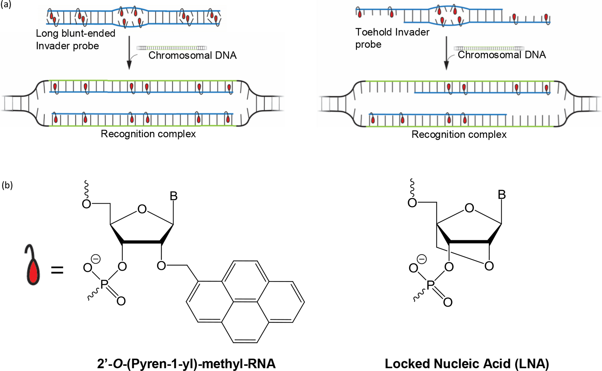
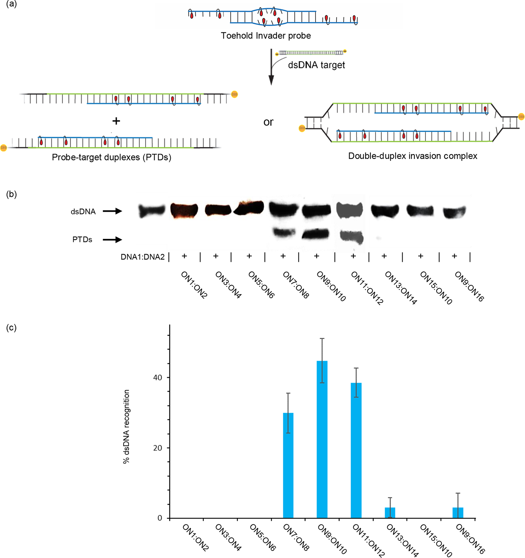
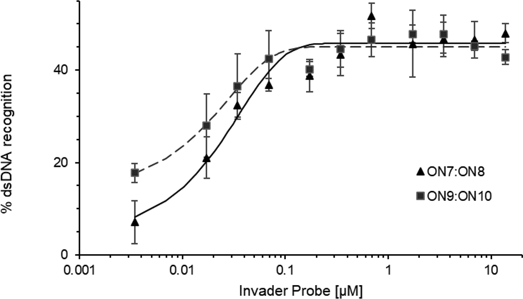
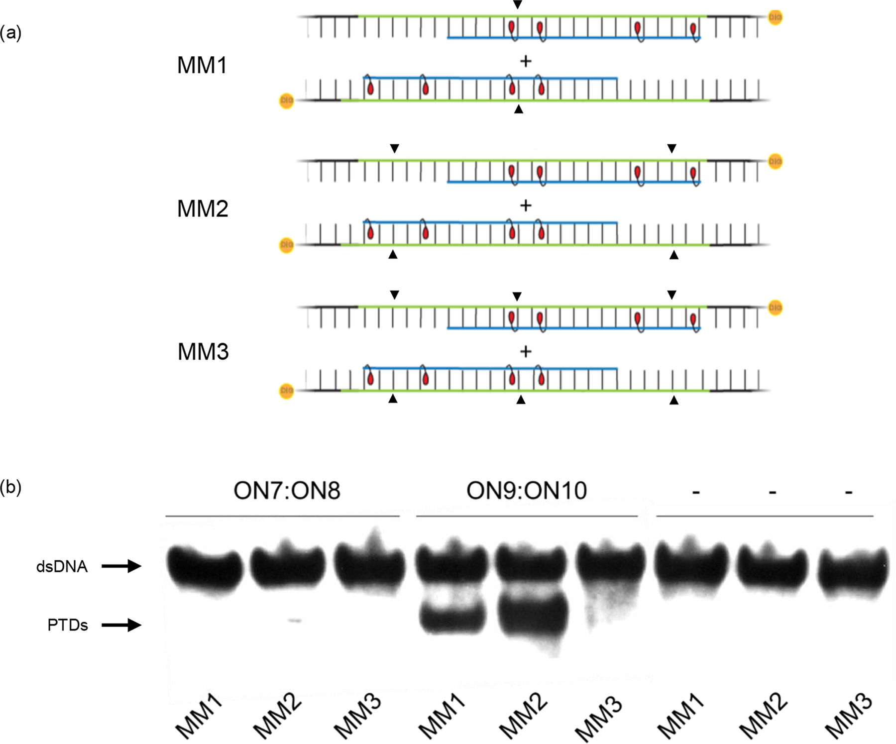
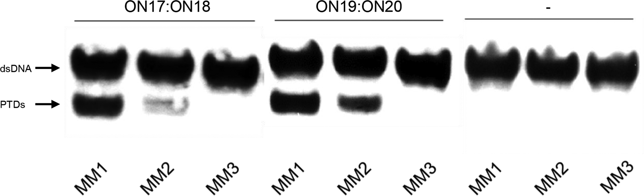
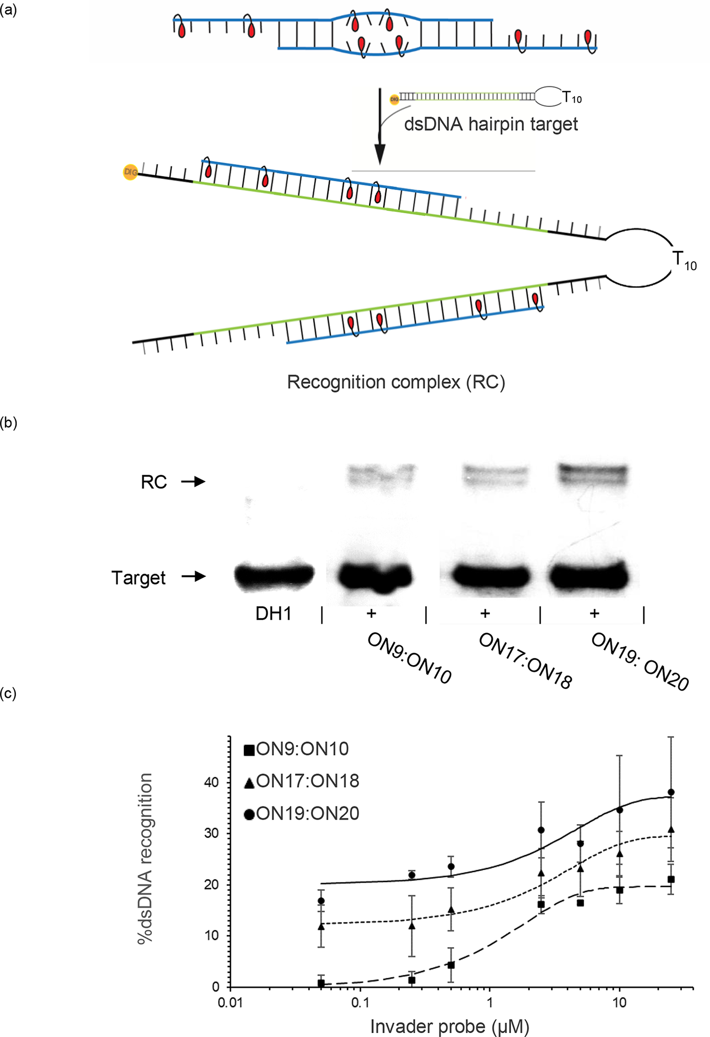
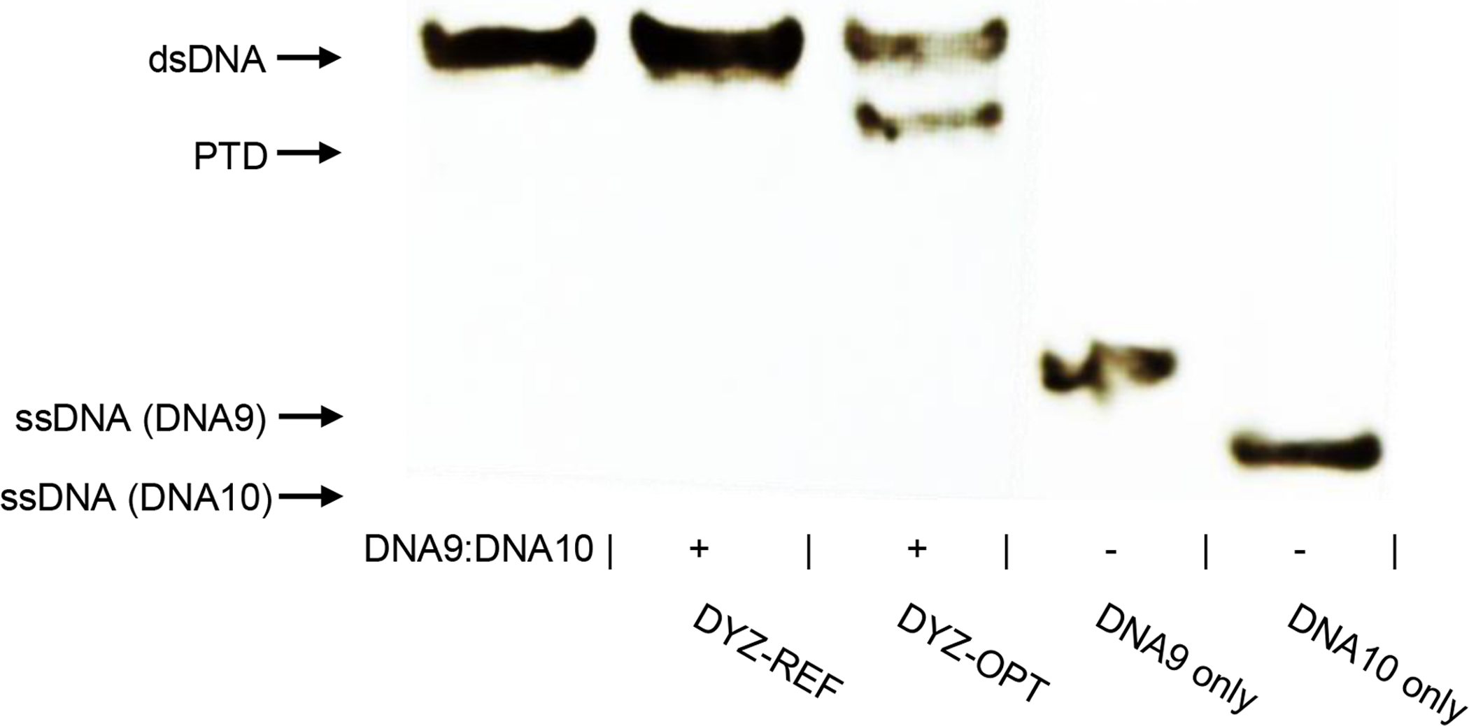
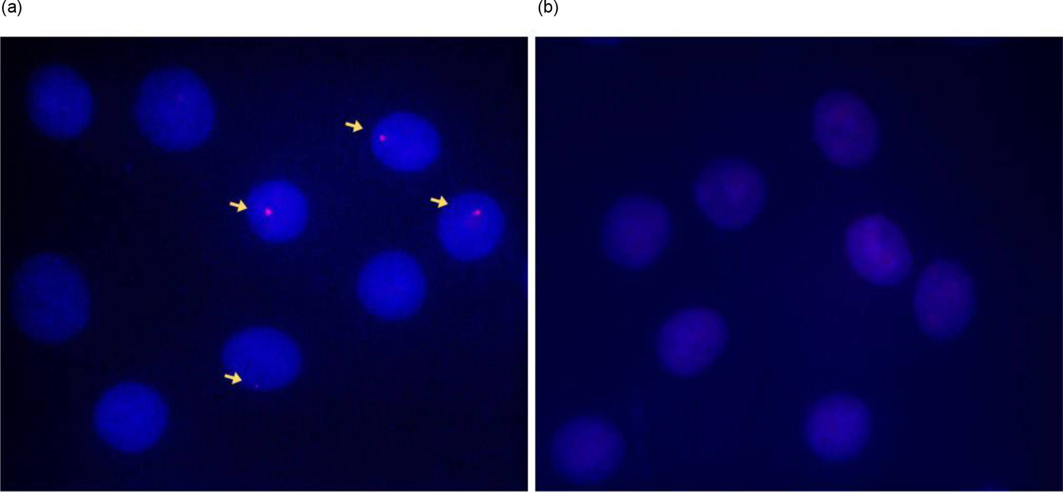
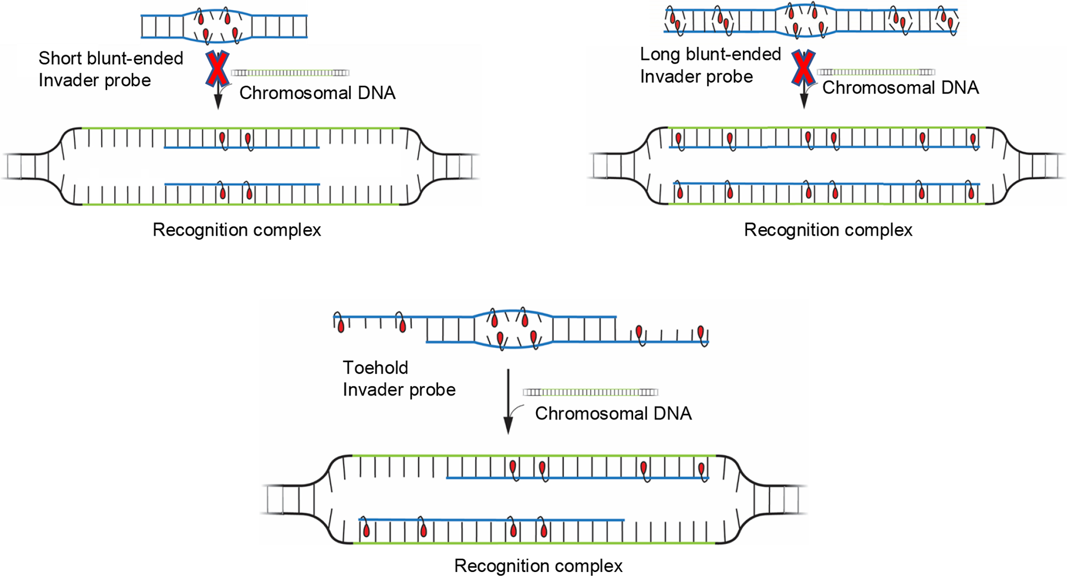
Development of molecules capable of binding to specific sequences of double-stranded (ds) DNA continues to attract considerable interest, as this may yield useful tools for applications in life science, biotechnology, and medicine. We have previously demonstrated sequence-unrestricted of dsDNA using Invader probes, i.e., DNA duplexes that are energetically activated through incorporation of +1 interstrand zipper arrangements of O2'-intercalator-functionalized RNA monomers. Nonetheless, recognition of extended dsDNA target regions remains challenging due to the high stability of the corresponding probes. To address this, we introduce toehold Invader probes, i.e., Invader probes with 5'-single-stranded overhangs. This design provides access to probes with shortened double-stranded segments, which facilitates probe denaturation. The single-stranded overhangs can, furthermore, be modified with affinity-enhancing modifications like LNA (locked nucleic acid) monomers to additionally increase target affinity. Herein, we report the biophysical and dsDNA-targeting properties of different toehold Invader designs and compare them to conventional Invader probes. LNA-modified toehold Invader probes display promising recognition characteristics, including greatly improved affinity to dsDNA, excellent binding specificity, and fast recognition kinetics, which enabled recognition of chromosomal DNA targets that have proven refractory to recognition by conventional Invader probes. Thus, toehold Invader probes represent another step toward a robust, oligonucleotide-based approach for sequence-unrestricted dsDNA-recognition.


Conflict of interest statement

CONFLICTS OF INTERESTS

P. J. H. is an inventor on patents pertaining to Invader probes, which have been issued to the University Idaho.

Figures

Figure 1.
Figure 1.
(a) Illustration of dsDNA-recognition principle using conventional and toehold Invader probes. (b) Structures of 2’-O-(pyren-1-yl)methyl-RNA and LNA monomers used herein.
Figure 2.
Figure 2.
(a) Illustration of assay used to evaluate Invader-mediated recognition of model dsDNA targets along with possible outcomes of the recognition process. (b) Representative gel electrophoretograms from recognition experiments in which DNA1:DNA2 was incubated with a 5-fold molar excess of different probes. PTDs = probe-target duplexes. (c) Histogram depicting averaged results from at least three independent recognition experiments with error bars representing standard deviation. Conditions: pre-annealed doubly 3’-DIG-labeled DNA1:DNA2 (50 nM) was incubated with a 5-fold molar excess of the specified pre-annealed probe in HEPES buffer (50 mM HEPES, 100 mM NaCl, 5 mM MgCl2, pH 7.2, 10% sucrose, 1.44 mM spermine tetrahydrochloride) at 37 °C for 17 h. Mixtures were resolved on 16% nd-PAGE gels.
Figure 3.
Figure 3.
Dose-response curves for recognition of DNA1:DNA2 by Invader probes ON7:ON8 and ON9:ON10 at 37 °C. Representative electrophoretograms are shown in Fig. S30†. Experimental conditions are as described in Fig. 2.
Figure 4.
Figure 4.
Binding specificity of toehold Invader probes. (a) Illustration of mismatched probe-target duplexes that would form if MM1-MM3 were recognized by ON9:ON10; arrowheads indicate the position of mismatched nucleotide. (b) Representative electrophoretograms from experiments in which a 5-fold excess of ON7:ON8 or ON9:ON10 was incubated with non-complementary MM1-MM3 targets (see Table S6† for sequences) as described in Figure 2. PTDs = probe-target duplexes.
Figure 5.
Figure 5.
Binding specificity of toehold Invader probes. Representative electrophoretograms from experiments in which a 5-fold molar excess of ON17:ON18 or ON19:ON20 was incubated with non-complementary targets MM1-MM3 at 37 °C for 17 h. For sequences of MM1-MM3, see Table S11†. Pre-annealed 3’-DIG-labeled MM1-MM3 (50 nM) was incubated with pre-annealed Invader probe at 37 °C for 17 h in HEPES buffer as outlined in Fig. 2. PTD = probe-target duplex.
Figure 6.
Figure 6.
(a) Assay used to evaluate recognition of DNA hairpin targets using LNA-modified toehold Invader probes. (b) Representative gel electrophoretogram from recognition experiments in which a 50-fold molar excess of toehold Invader probes was incubated with DNA hairpin DH1 (50 nM). (c) Dose-response curves for recognition of DH1 using toehold Invader probes ON9:ON10, ON17:ON18, or ON19:ON20. Error bars represent standard deviation from at least three experiments. Experimental conditions are as stated in Fig. 2, with the exception of using 12% nd-PAGE gels.
Figure 7.
Figure 7.
Representative gel electrophoretograms from recognition experiments in which pre-annealed 3’-DIG-labeled dsDNA target DNA9:DNA10 (50 nM) was incubated with a 5-fold molar excess of DYZ-REF or DYZ-OPT as outlined in Fig. 2. DYZ-REF = 5’-Cy3-TUAUATGCTGUTCTC:3’-AAUAUACGACAAGAG-Cy3. DYZ-OPT = 5’-Cy3-TgTgTG-TUAUATGCTGUTCTC:3’-AAUAUACGACAAGAG-TCgGgA-Cy3. Modifications are indicated as described in Table 1. DNA9 = 5’-TGACTGTGTGTTATATGCTGTTCTCAGCCCTTGAC. DNA10 = 3’-ACTGACACACAATATACGACAAGAGTCGGGAACTG.
Figure 8.
Figure 8.
Images from FISH experiments using toehold Invader probes (a) DYZ-OPT (3 ng), and (b) DYZ-REF (15 ng) under non-denaturing conditions. Fixed isolated nuclei from male bovine kidney cells were incubated with probes for 3 h at 37.5 °C in a Tris buffer (20 mM Tris-Cl, 100 mM KCl, pH 8.0) and counterstained with DAPI. Images were obtained by overlaying images from Cy3 (red) and DAPI (blue) channels and adjusting the exposure. Nuclei were viewed at 60X magnification using a Nikon Eclipse Ti-S inverted microscope. The probe amounts used were chosen based on initial optimization studies (Fig. S42†).
Figure 9.
Figure 9.
Comparison of different Invader probe architectures.

Similar articles

Cited by

References

    1. Hsu PD, Lander ES and Zhang F, Cell, 2014, 157, 1262–1278. - PMC - PubMed
    1. Komor AC, Badran AH and Liu DR, Cell, 2017, 168, 20–36. - PMC - PubMed
    1. Duca M, Vekhoff P, Oussedik K, Halby L and Arimondo PB, Nucleic Acids Res, 2008, 36, 5123–5138. - PMC - PubMed
    1. Hari Y, Obika S and Imanishi T, Eur. J. Org. Chem, 2012, 2875–2887.
    1. Kaihatsu K, Janowski BA and Corey DR, Chem. Biol, 2004, 11, 749–758. - PubMed

Publication types

MeSH terms