. 2021 Dec;33(51):e2106155.
doi: 10.1002/adma.202106155. Epub 2021 Oct 17.

Plasmonic Amyloid Tactoids

Affiliations

Plasmonic Amyloid Tactoids

Ye Yuan et al. Adv Mater. 2021 Dec.

Abstract

Despite their link to neurodegenerative diseases, amyloids of natural and synthetic sources can also serve as building blocks for functional materials, while possessing intrinsic photonic properties. Here, it is demonstrated that orientationally ordered amyloid fibrils exhibit polarization-dependent fluorescence, and can mechanically align rod-shaped plasmonic nanoparticles codispersed with them. The coupling between the photonic fibrils in liquid crystalline phases and the plasmonic effect of the nanoparticles leads to selective activation of plasmonic extinctions as well as enhanced fluorescence from the hybrid material. These findings are consistent with numerical simulations of the near-field plasmonic enhancement around the nanoparticles. The study provides an approach to synthesize the intrinsic photonic and mechanical properties of amyloid into functional hybrid materials, and may help improve the detection of amyloid deposits based on their enhanced intrinsic luminescence.

Keywords: amyloid fibrils; fluorescence; gold nanorods; liquid crystals; plasmonics; self-assembly.


Conflict of interest statement

The authors declare no conflict of interest.

Figures

Figure 1
Figure 1
Polarization‐dependent fluorescence of amyloid fibrils tactoids. a) Polarizing microscopy image of bipolar tactoids. The white double arrows represent the crossed polarizers. b) Corresponding fluorescence images taken at different sample orientation with respect to the fixed polarization of the excitation light. c,d) Polarizing and fluorescence microscopy images of a cholesteric tactoid. All the fluorescence images are obtained with a 405 nm excitation laser and the linear laser polarization is marked with P. e–g) Polarization‐dependent fluorescence spectra measured within and outside of tactoids using 405 nm (e), 514 nm (f), and 633 nm (g) excitation light. Colored lines are spectra obtained with the polarization of excitation parallel to the fibril orientation and black lines are spectra obtained with the polarization of excitation perpendicular to the fibril orientation. The solid lines represent the spectra obtained within tactoids, while the dotted lines represent the spectra obtained outside of tactoids. See Figure S1, Supporting Information for the corresponding fluorescence images.
Figure 2
Figure 2
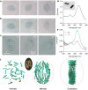
Amyloid fibril (AF)‐gold nanorod (GNR) hybrid tactoids. a) Microscopy images showing the color of a hybrid tactoid rotated under unpolarized illumination light. b) The same tactoid with a fixed polarization of illumination light (marked with P). c) Microscopy images of a cholesteric hybrid tactoid under linearly polarized light. d) Extinction spectrum of PEG‐capped GNRs dispersed in water. The inset shows an SEM image of the GNRs used. e) Polarization‐dependent extinction spectra of a hybrid tactoid. See the corresponding microscopy images in Figure S5 (Supporting Information). f) Schematics of AF‐GNR codispersion in isotropic, nematic, and cholesteric phases. The latter two are shown in bipolar and cholesteric tactoids. The long green rods represent the AFs and the short golden rods represent the GNRs; the schematics are not true to scale. The inset is a 3D reconstruction of a bipolar hybrid tactoid from a stack of fluorescence images as in Figure S4, Supporting Information.
Figure 3
Figure 3
Plasmonic enhanced fluorescence of AF‐GNR hybrid tactoids. a) Fluorescence image of hybrid tactoids when the polarization of the excitation light (633 nm) is parallel to the long axis of a bipolar tactoid. Inset at the bottom shows the image obtained in transmission mode. b) Fluorescence and transmission microscopy images of the tactoids in (a) rotated 90°. c–e) Polarization dependent fluorescence spectra measured within and outside of a hybrid tactoid (marked with the red arrows in (a) and (b) using 633 nm (c), 514 nm (d), and 405 nm (e) excitation light. The lines are color‐coded and dashed the same way as in Figure 1.
Figure 4
Figure 4
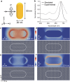
Numerical simulations of plasmonic responses of GNRs. a) Schematic showing the parameters and setup for the simulations. The dimensions of the GNRs are 20 × 50 nm and plane‐wave incident light with varying polarizations is used as the excitation. b) Simulated and experimental extinction spectra of GNRs dispersed in water. c–f) Simulated near‐field enhancement of a GNR excited with 633 and 514 nm light. The colors represent the order of magnitude of electric field enhancement, i.e., log(E 2/E 0 2); note the different maximum values when excited with 633 and 514 nm. The incident light propagates along z axis as illustrated in (a) and the ensuing field distribution is extracted as the central cross‐sections in the xy plane. The insets at the bottom of each panel are the simulated distribution of polarization with the white dashes representing the local direction of polarization; the capsule shapes in the insets indicate the boundary of the GNR. The size of the simulated areas is 40 × 100 nm2; the incident polarization P, defined along the direction of the electric field, is marked with white double arrows.

References

    1. Dobson C. M., Nature 2003, 426, 884. - PubMed
    1. Eisenberg D., Jucker M., Cell 2012, 148, 1188. - PMC - PubMed
    1. Sawaya M. R., Sambashivan S., Nelson R., Ivanova M. I., Sievers S. A., Apostol M. I., Thompson M. J., Balbirnie M., Wiltzius J. J., McFarlane H. T., Madsen A. O., Riekel C., Eisenberg D., Nature 2007, 447, 453. - PubMed
    1. Wasmer C., Lange A., Van Melckebeke H., Siemer A. B., Riek R., Meier B. H., Science 2008, 319, 1523. - PubMed
    1. Smith J. F., Knowles T. P. J., Dobson C. M., MacPhee C. E., Welland M. E., Proc. Natl. Acad. Sci. USA 2006, 103, 15806. - PMC - PubMed

LinkOut - more resources