. 2021 Sep 30:12:709123.
doi: 10.3389/fphys.2021.709123. eCollection 2021.

MicroRNA-195-5p Downregulation Inhibits Endothelial Mesenchymal Transition and Myocardial Fibrosis in Diabetic Cardiomyopathy by Targeting Smad7 and Inhibiting Transforming Growth Factor Beta 1-Smads-Snail Pathway

Affiliations

MicroRNA-195-5p Downregulation Inhibits Endothelial Mesenchymal Transition and Myocardial Fibrosis in Diabetic Cardiomyopathy by Targeting Smad7 and Inhibiting Transforming Growth Factor Beta 1-Smads-Snail Pathway

Huaisheng Ding et al. Front Physiol. .

Abstract

Diabetic cardiomyopathy (DCM) is a complication of diabetes mellitus, which is associated with fibrosis and microRNAs (miRs). This study estimated the mechanism of miR-195-5p in endothelial mesenchymal transition (EndMT) and myocardial fibrosis in DCM. After the establishment of DCM rat models, miR-195-5p was silenced by miR-195-5p antagomir. The cardiac function-related indexes diastolic left ventricular anterior wall (LVAW, d), systolic LVAW (d), diastolic left ventricular posterior wall (LVPW, d), systolic LVPW (d), left ventricular ejection fraction (LVEF), and fractional shortening (FS) were measured and miR-195-5p expression in myocardial tissue was detected. Myocardial fibrosis, collagen deposition, and levels of fibrosis markers were detected. Human umbilical vein endothelial cells (HUVECs) were exposed to high glucose (HG) and miR-195-5p was silenced. The levels of fibrosis proteins, endothelial markers, fibrosis markers, EndMT markers, and transforming growth factor beta 1 (TGF-β1)/Smads pathway-related proteins were measured in HUVECs. The interaction between miR-195-5p and Smad7 was verified. In vivo, miR-195-5p was highly expressed in the myocardium of DCM rats. Diastolic and systolic LVAW, diastolic and systolic LVPW were increased and LVEF and FS were decreased. Inhibition of miR-195-5p reduced cardiac dysfunction, myocardial fibrosis, collagen deposition, and EndMT, promoted CD31 and VE-cadehrin expressions, and inhibited α-SMA and vimentin expressions. In vitro, HG-induced high expression of miR-195-5p and the expression changes of endothelial markers CD31, VE-cadehrin and fibrosis markers α-SMA and vimentin were consistent with those in vivo after silencing miR-195-5p. In mechanism, miR-195-5p downregulation blocked EndMT by inhibiting TGF-β1-smads pathway. Smad7 was the direct target of miR-195-5p and silencing miR-195-5p inhibited EndMT by promoting Smad7 expression. Collectively, silencing miR-195-5p inhibits TGF-β1-smads-snail pathway by targeting Smad7, thus inhibiting EndMT and alleviating myocardial fibrosis in DCM.

Keywords: TGF-β1-smads-snail pathway; diabetic cardiomyopathy; endothelial mesenchymal transition; miR-195-5p; myocardial fibrosis; smad7.


Conflict of interest statement

The authors declare that the research was conducted in the absence of any commercial or financial relationships that could be construed as a potential conflict of interest.

Figures

Figure 1
Figure 1
Inhibition of microRNA (miR)-195-5p reduces cardiac dysfunction in diabetic cardiomyopathy (DCM) rats. (A) Expression changes of rats in each group were detected using reverse transcription quantitative PCR (RT-qPCR); (B) Blood glucose changes of rats in each group; (C) Insulin level changes of rats in each group; (D) E/A ratio of rats in each group; (E) SRe/SRa ratio of rats in each group; (F) Relative diastolic left ventricular anterior wall (LVAW) in rats; (G) Relative systolic LVAW in rats; (H) Relative diastolic left ventricular posterior wall (LVPW) in rats; (I) Relative systolic LVPW in rats; (J) Relative left ventricular ejection fraction (LVEF) value in rats; and (K) Relative fractional shortening (FS) value in rats; N=12 rats in each group; the data were expressed as mean±SD, and analyzed by one-way ANOVA and Tukey’s multiple comparisons test; *p<0.05, ***p<0.001. d, diastole; s, systole.
Figure 2
Figure 2
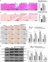
Inhibition of miR-195-5p reduces myocardial fibrosis in DCM rats. (A) Masson staining, scale bar=50μm, 200 ×; (B) Sirius red standing, scale bar=50μm, 200 ×; (C): Immunohistochemistry was adopted to detect the expression of collagen I, collagen III, fibronectin, and MMP-9; and (D) Western blot was used to detect the levels of collagen I, collagen III, fibronectin, and MMP-9. N=12 rats in each group; the data were expressed as mean±SD, and analyzed by one-way ANOVA and Tukey’s multiple comparisons test; *p<0.05, ***p<0.001.
Figure 3
Figure 3
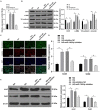
Inhibition of miR-195-5p can reduce the endothelial mesenchymal transition (EndMT) in DCM rats. (A) Western blot was used to detect the levels of CD31, α-SMA, VE-cadherin, and vimentin; (B) Immunofluorescence was used to detect the expression of CD31 and α-SMA in myocardial tissue, scale bar=25μm, 400 ×; (C) RT-qPCR was used to detect the expression of snail and twist; and (D) Western blot was used to detect the levels of snail and twist; N=12 rats in each group; the data were expressed as mean±SD, and analyzed by one-way ANOVA and Tukey’s multiple comparisons test; *p<0.05, ***p<0.001.
Figure 4
Figure 4
Inhibition of miR-195-5p attenuates high glucose (HG)-induced EndMT in human umbilical vein endothelial cells (HUVECs). (A) RT-qPCR was used to detect the expression of miR-195-5p; (B) Western blot was used to detect the levels of CD31, α-SMA, VE-cadherin, and vimentin; (C) Immunofluorescence was used to detect the expression of VE-cadherin and vimentin in endothelial cells (scale bar=100μm, 100 ×); (D) RT-qPCR was used to detect the expression of snail and twist; and (E) Western blot was used to detect the levels of snail and twist; the cell experiment was repeated three times. The data were expressed as mean±SD, and analyzed by one-way ANOVA and Tukey’s multiple comparisons test; *p<0.05.
Figure 5
Figure 5
Inhibition of miR-195-5p inhibits EndMT by inhibiting transforming growth factor beta 1 (TGF-β1)-smads pathway. (A) Western blot was used to detect the levels of TGF-β1, Smad2/3, p-Smad2/3, and smad7 in myocardial tissue; (B) Western blot was used to detect the levels of TGF-β1, Smad2/3, p-Smad2/3, and smad7 in endothelial cells; (C) RT-qPCR was used to detect the expression of miR-195-5p in endothelial cells; (D) Western blot was used to detect the levels of TGF-β1, Smad2/3, p-Smad2/3, and smad7 after LY-364947 treatment; (E) Western blot was used to detect the levels of CD31, α-SMA, VE-cadherin, and vimentin; (F) Immunofluorescence was used to detect the expression of VE cadherin and vimentin, scale bar=100μm, 100 ×; the cell experiment was repeated three times. The data were expressed as mean±SD, and analyzed by one-way ANOVA and Tukey’s multiple comparisons test; *p<0.05.
Figure 6
Figure 6
miR-195-5p can bind to smad7. (A) TargetScan (http://www.targetscan.org/vert_71/) predicted the binding of miR-195-5 and smad7; (B) Dual-luciferase assay showed that miR-195-5 could bind smad7; and (C) RNA immunoprecipitation (RIP) assay showed that miR-195-5 could bind to smad7; the cell experiment was repeated three times. The data were expressed as mean±SD, and analyzed by independent t test; *p<0.05.
Figure 7
Figure 7
Silencing miR-195-5p inhibits EndMT by promoting smad7 expression. (A) The expression of miR-195-5p and smad7 was detected by RT-qPCR; (B) The levels of CD31, α-SMA, VE-cadherin, and vimentin was detected by Western blot; (C) The expression of VE-cadherin and vimentin was detected by immunofluorescence, scale bar=100μm, 100 ×; (D) The expression of snail and twist was detected by RT-qPCR; and (E) Western blot was used to detect the levels of snail and twist; the cell experiment was repeated three times. The data were expressed as mean±SD, and analyzed by one-way ANOVA and Tukey’s multiple comparisons test; *p<0.05.

Similar articles

Cited by

References

    1. Becirovic-Agic M., Chalise U., Daseke M. J., Konfrst S., Salomon J. D., Mishra P. K., et al. . (2021). Infarct in the heart: what's MMP-9 got to do with it? Biomolecules 11:491. doi: 10.3390/biom11040491, PMID: - DOI - PMC - PubMed
    1. Bugger H., Abel E. D. (2014). Molecular mechanisms of diabetic cardiomyopathy. Diabetologia 57, 660–671. doi: 10.1007/s00125-014-3171-6, PMID: - DOI - PMC - PubMed
    1. Cao Y., Feng B., Chen S., Chu Y., Chakrabarti S. (2014). Mechanisms of endothelial to mesenchymal transition in the retina in diabetes. Invest. Ophthalmol. Vis. Sci. 55, 7321–7331. doi: 10.1167/iovs.14-15167, PMID: - DOI - PubMed
    1. Chakraborty C., Doss C. G., Bandyopadhyay S., Agoramoorthy G. (2014). Influence of miRNA in insulin signaling pathway and insulin resistance: micro-molecules with a major role in type-2 diabetes. Wiley Interdiscip. Rev. RNA 5, 697–712. doi: 10.1002/wrna.1240, PMID: - DOI - PubMed
    1. Che H., Wang Y., Li Y., Lv J., Li H., Liu Y., et al. . (2020). Inhibition of microRNA-150-5p alleviates cardiac inflammation and fibrosis via targeting Smad7 in high glucose-treated cardiac fibroblasts. J. Cell. Physiol. 235, 7769–7779. doi: 10.1002/jcp.29386, PMID: - DOI - PubMed