. 2021 Sep 30:12:712509.
doi: 10.3389/fphys.2021.712509. eCollection 2021.

Mechanical Strain Regulates Myofibroblast Differentiation of Human Scleral Fibroblasts by YAP

Affiliations

Mechanical Strain Regulates Myofibroblast Differentiation of Human Scleral Fibroblasts by YAP

Di Hu et al. Front Physiol. .

Abstract

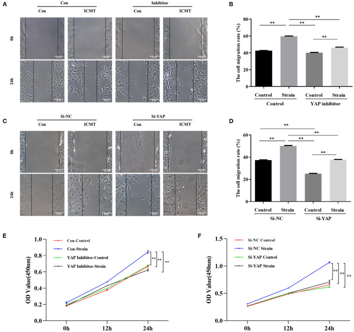
Scleral extracellular matrix (ECM) remodeling is thought to play a critical role in the pathogenesis of glaucoma. Mechanical strain induced by elevated intraocular pressure can promote myofibroblast differentiation of fibroblasts and result in scleral ECM remodeling; however, the underlying mechanism remains poorly understood. Yes-associated protein (YAP) is a mechanosensory protein and the key downstream transcriptional effector of the Hippo signaling pathway. Here, we investigated the role of YAP in mechanical strain-induced myofibroblast transformation during glaucoma scleral ECM remodeling. Integrative bioinformatics analyses were performed to identify the key pathways for the ECM remodeling of the sclera in glaucoma. Sprague-Dawley rats were used to establish a chronic ocular hypertension model, and the expression of collagen type I (COL1) and YAP in the sclera was analyzed by immunohistochemical analysis and Western blotting. Furthermore, human scleral fibroblasts (HSFs) were cultured and subjected to mechanical strain. In groups with or without the YAP siRNA or YAP inhibitor, cell proliferation, migration capacity, and the expression levels of YAP, COL1, and α-smooth muscle actin (α-SMA) were evaluated by Cell Counting Kit-8 assay, scratch assay, and Western blotting. The interactions between YAP and Smad3 were demonstrated by coimmunoprecipitation, and the expression levels of COL1 and α-SMA were evaluated in groups treated with or without the Smad3 inhibitor. We first revealed that the Hippo signaling pathway may be involved in mechanical strain-induced scleral ECM remodeling through bioinformatics analysis. Furthermore, the in vivo study showed upregulated YAP, COL1, and α-SMA expression in the hypertensive sclera of rats. In vitro, mechanical strain increased YAP and COL1 expression in HSFs and promoted myofibroblast differentiation. After YAP knockdown or inhibition with verteporfin, mechanical strain-induced fibrotic changes in HSFs were markedly suppressed. Additionally, YAP showed a protein interaction with Smad3, and the upregulation of a-SMA and COL1 in response to mechanical strain was also significantly downregulated following the inhibition of Smad3. In conclusion, mechanical strain activated scleral myofibroblast differentiation via YAP. The YAP pathway may play an important role in regulating scleral myofibroblast differentiation and ECM remodeling of the sclera in glaucoma.

Keywords: extracellular matrix remodeling; glaucoma; mechanical strain; myofibroblast differentiation; sclera.


Conflict of interest statement

The authors declare that the research was conducted in the absence of any commercial or financial relationships that could be construed as a potential conflict of interest.

Figures

Figure 1
Figure 1
Key signaling pathway in mechanical strain-induced scleral ECM remodeling in glaucoma. (A) Overall bioinformatics analysis strategy. (B) Venn diagram of the text mining genes related to glaucoma, sclera, mechanical strain, and EMC remodeling. The 88 common genes were considered to be related to mechanical strain-induced scleral ECM remodeling in glaucoma. (C) The protein-protein interaction (PPI) network of the 54 target text mining genes, red circles represent the significant module genes. (D) Significantly enriched KEGG signaling pathways in gene module. Abbreviations: EMC, extracellular matrix; KEGG, Kyoto Encyclopedia of Genes and Genomes.
Figure 2
Figure 2
Yap expression in the OHT model of SD rats. (A) Intraocular pressure (IOP) in the OHT model of SD rats. (B) Representative picture and quantitative analysis of immunofluorescent staining of COL1 in the OHT model of SD rats (scale bar = 50 um). (C) Representative pictures and quantitative analysis of immunofluorescent staining of YAP in the OHT model of SD rats (scale bar = 20 um). (D) Western blotting analyses of YAP in the OHT model of SD rats. (E) Representative picture of immunofluorescence of containing YAP and α-SMA in the OHT model of SD rats (scale bar = 10 um). (F) Quantitative analysis of immunofluorescence of YAP and α-SMA in the OHT model of SD rats. Scleral tissues were collected after the models were successfully established for 1 week. The results are presented as means ± SD (n = 3), *P < 0.05. Abbreviations: OHT, ocular hypertension; SD, Sprague–Dawley; NT, normotension; COL1, collagen type I; GAPDH, glyceraldehyde 3-phosphate dehydrogenase; α-SMA, α-smooth muscle actin; DAPI, 4′,6-diamidino-2-phenylindole.
Figure 3
Figure 3
YAP is involved in the regulation of myofibroblast differentiation induced by the mechanical strain. (A) Western blotting and quantitative analysis of YAP, COL1, and α-SMA under mechanical strain. HSF cultures without applying strain at 8 h served as control groups. HSFs of the strain group were subjected to 10% cyclic strain at 0.5 Hz for 4, 8, 12, and 24 h. (B) Western blotting and quantitative analyses of YAP, COL1, and α-SMA in the YAP-specific siRNA and strain treatment groups. HSFs were transfected with the control siRNA (Con) or YAP siRNA (siRNA). HSFs of the strain group were subjected to 10% cyclic strain at 0.5 Hz for 8 h. (C) Western blotting and quantitative analyses of YAP, COL1, and α-SMA in the YAP-specific inhibitor and strain treatment groups. HSFs were transfected without (Con) or with verteporfin at 75 mg/ml (inhibitor). HSFs of the strain group were subjected to 10% cyclic strain at 0.5 Hz for 8 h. Data are presented as means ± SD of three separate experiments. **P < 0.001. Abbreviations: Con, control; ICMT, intermittent cyclic mechanical tension; α-SMA, α-smooth muscle actin; COL1, collagen type I; GAPDH, glyceraldehyde 3-phosphate dehydrogenase.
Figure 4
Figure 4
Mechanical strain induces HSFs motility and proliferation by activating YAP. (A) HSFs motility was detected by the scratch assay in the context of YAP inhibition. (B) Quantitative analysis of cell migration. (C) HSFs motility was detected by the scratch assay in the context of YAP knockdown. (D) Quantitative analysis of cell migration. (E) HSFs proliferative capacity was determined by CCK-8 assay in the context of YAP inhibition. (F) HSFs proliferative capacity was determined by CCK-8 assay in the context of YAP knockdown. HSFs of the strain group were subjected to 10% cyclic strain at 0.5 Hz for 8 h. **P < 0.001. Con, control; ICMT, intermittent cyclic mechanical tension.
Figure 5
Figure 5
Smad3 was bound to YAP involved in myofibroblast differentiation induced by the mechanical strain. (A) Immunoblot of the coimmunoprecipitation was performed to test if YAP binds to Smad3. HSF cultures without applying strain at 8 h served as control groups. HSFs of the strain group were subjected to 10% cyclic strain at 0.5 Hz for 8 h. (B) Western blotting and quantitative analyses of COL1 and α-SMA in the Smad3-specific inhibitor and strain treatment groups. HSFs were transfected without (Con) or with SIS3 at 10 μM (inhibitor). HSFs of the strain group were subjected to 10% cyclic strain at 0.5 Hz for 8 h. Data are presented as means ± SD of three separate experiments. *P < 0.05. Abbreviations: Con, control; ICMT, intermittent cyclic mechanical tension; IP, immunoprecipitation; α-SMA, α-smooth muscle actin; COL1, collagen type I; GAPDH, glyceraldehyde 3-phosphate dehydrogenase.

Similar articles

Cited by

References

    1. Anderson D. R., Drance S. M., Schulzer M. (2003). Factors that predict the benefit of lowering intraocular pressure in normal tension glaucoma. Am. J. Ophthalmol. 136, 820–829. 10.1016/S0002-9394(03)00478-1 - DOI - PubMed
    1. Baum J., Duffy H. S. (2011). Fibroblasts and myofibroblasts: what are we talking about? J. Cardiovasc. Pharmacol. 57, 376–379. 10.1097/FJC.0b013e3182116e39 - DOI - PMC - PubMed
    1. Bellezza A. J., Rintalan C. J., Thompson H. W., Downs J. C., Hart R. T., Burgoyne C. F. (2003). Deformation of the lamina cribrosa and anterior scleral canal wall in early experimental glaucoma. Invest. Ophthalmol. Vis. Sci. 44, 623–637. 10.1167/iovs.01-1282 - DOI - PubMed
    1. Bergmeier V., Etich J., Pitzler L., Frie C., Koch M., Fischer M., et al. . (2018). Identification of a myofibroblast-specific expression signature in skin wounds. Matrix Biol. 65, 59–74. 10.1016/j.matbio.2017.07.005 - DOI - PubMed
    1. Boote C., Sigal I. A., Grytz R., Hua Y., Nguyen T. D., Girard M. J. A. (2020). Scleral structure and biomechanics. Prog. Retin. Eye Res. 74:100773. 10.1016/j.preteyeres.2019.100773 - DOI - PMC - PubMed

LinkOut - more resources