Review
. 2021 Sep 29:12:715527.
doi: 10.3389/fphys.2021.715527. eCollection 2021.

The Manifold Roles of Sphingolipids in Viral Infections

Affiliations
Review

The Manifold Roles of Sphingolipids in Viral Infections

Elita Avota et al. Front Physiol. .

Abstract

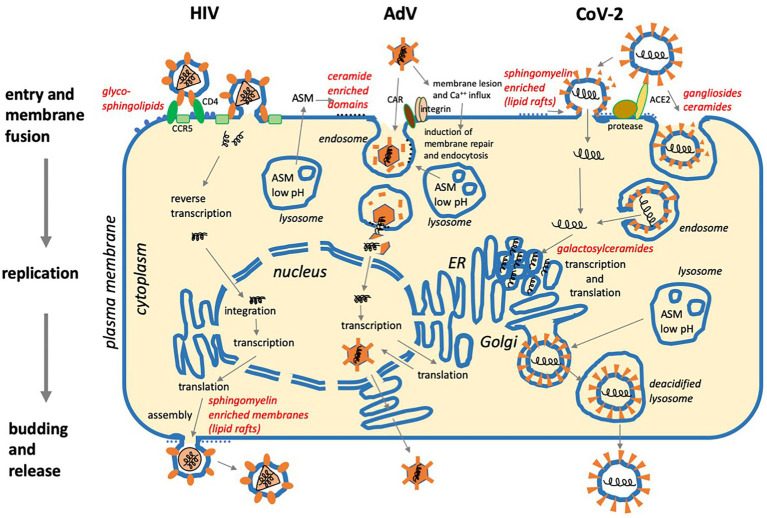
Sphingolipids are essential components of eukaryotic cells. In this review, we want to exemplarily illustrate what is known about the interactions of sphingolipids with various viruses at different steps of their replication cycles. This includes structural interactions during entry at the plasma membrane or endosomal membranes, early interactions leading to sphingolipid-mediated signal transduction, interactions with internal membranes and lipids during replication, and interactions during virus assembly and budding. Targeted interventions in sphingolipid metabolism - as far as they can be tolerated by cells and organisms - may open novel possibilities to support antiviral therapies. Human immunodeficiency virus type 1 (HIV-1) infections have intensively been studied, but for other viral infections, such as influenza A virus (IAV), measles virus (MV), hepatitis C virus (HCV), dengue virus, Ebola virus, and severe acute respiratory syndrome coronavirus type 2 (SARS-CoV-2), investigations are still in their beginnings. As many inhibitors of sphingolipid metabolism are already in clinical use against other diseases, repurposing studies for applications in some viral infections appear to be a promising approach.

Keywords: ceramide; plasma membrane; sphingolipid; sphingosine-1-phosphate; virus budding; virus entry; virus replication.


Conflict of interest statement

The authors declare that the research was conducted in the absence of any commercial or financial relationships that could be construed as a potential conflict of interest.

Figures

Figure 1
Figure 1
(A) Schematic representation of the sphingolipid metabolism from de novo synthesis from palmitoyl-CoA and serine to the final degradation to phosphoethanolamine and hexadecenal. Involved enzymes are neutral and acid sphingomyelinase (ASM; neutral sphingomyelinase, NSM), sphingomyelin synthetase (SMS), ceramidase, sphingosine kinases 1 and 2 (SphK-1,2), and sphingosine-1-phosphate (S1P) lyase. (B) Examples of the chemical structures of sphingolipids with sphingomyelin (SM) and ceramides of chain length 16 (C16). (C) Characteristic ratios of amounts of SM, ceramide (Cer), sphingosine, and sphingosine-1-phosphate (S1) in primary human peripheral blood lymphocytes (PBL; example form our own research).
Figure 2
Figure 2
Three examples of viral replication cycles and involvement of sphingolipids: (1) Human immunodeficiency virus (HIV) binds to glycoshingolipids, CD4, and co-receptors CCR5 or CXCR4 prior to membrane fusion at the plasma membrane. After reverse transcription of the positive-strand RNA genome and integration in the cellular DNA, the virus exploits the cellular machinery for transcription and translation. Assembly and budding take place at the plasma membrane with the involvement of sphingomyelin-enriched membrane domains (lipid rafts). (2) Adenovirus (AdV) binds to CAR and integrins, then causes membrane lesions which induce Ca++ influx and membrane repair. This occurs with the help of lysosomes requiring ASM activity, which supports viral uptake by endocytosis. After the disintegration of the viral capsid, the viral DNA is transported to the nucleus, and after transcription and translation, viral particles are assembled in the nucleus and released. (3) SARS Coronavirus-2 (CoV-2) requires protease activity and binds to ACE2, which is supported by sialic acid in ganglioside-enriched membrane domains prior to cell entry via fusion at the plasma membrane or endocytosis and subsequent membrane fusion. Transcription and translation take place on the cytoplasmic face of the ER. Galactosylceramide supports viral replication. After assembly, virus particles traffic to lysosomes for egress via lysosomal exocytosis (For references, see main text).
Figure 3
Figure 3
Schematic representation of a number of sphingolipid-associated signaling pathways and interactions with cellular functions potentially supporting or inhibiting viral replication. NSM, neutral sphingomyelinase; ASM, acid sphingomyelinase; SphK-1,2, sphingosine kinases 1 and 2; S1P, sphingosine-1-phosphate; CAPK, ceramide activated protein kinase; PKC, protein kinase C; PP2A, protein phosphatase 2 A; TRAF2, tumor necrosis factor associated factor 2; GRP94, glucose-regulated protein 94; IRE1a, inositol requiring enzyme 1 alpha; Hsp90, heat shock protein 90; RIP1, receptor-interacting protein 1; HDAC, histone deacetylase; and HIF-1a, hypoxia inducing factor 1 alpha.

References

    1. Aerts J., Artola M., van Eijk M., Ferraz M. J., Boot R. G. (2019). Glycosphingolipids and infection. Potential new therapeutic avenues. Front. Cell Dev. Biol. 7:324. doi: 10.3389/fcell.2019.00324, PMID: - DOI - PMC - PubMed
    1. Alfsen A., Bomsel M. (2002). HIV-1 gp41 envelope residues 650-685 exposed on native virus act as a lectin to bind epithelial cell galactosyl ceramide. J. Biol. Chem. 277, 25649–25659. doi: 10.1074/jbc.M200554200, PMID: - DOI - PubMed
    1. Aligeti M., Roder A., Horner S. M. (2015). Cooperation between the hepatitis C virus p7 and NS5B proteins enhances virion infectivity. J. Virol. 89, 11523–11533. doi: 10.1128/JVI.01185-15, PMID: - DOI - PMC - PubMed
    1. Alvarez S. E., Harikumar K. B., Hait N. C., Allegood J., Strub G. M., Kim E. Y., et al. . (2010). Sphingosine-1-phosphate is a missing cofactor for the E3 ubiquitin ligase TRAF2. Nature 465, 1084–1088. doi: 10.1038/nature09128, PMID: - DOI - PMC - PubMed
    1. Alvisi G., Madan V., Bartenschlager R. (2011). Hepatitis C virus and host cell lipids: an intimate connection. RNA Biol. 8, 258–269. doi: 10.4161/rna.8.2.15011, PMID: - DOI - PubMed