Clinical Trial
. 2021 Sep 30:12:732667.
doi: 10.3389/fimmu.2021.732667. eCollection 2021.

Poor CD4+ T Cell Immunogenicity Limits Humoral Immunity to P. falciparum Transmission-Blocking Candidate Pfs25 in Humans

Affiliations
Clinical Trial

Poor CD4+ T Cell Immunogenicity Limits Humoral Immunity to P. falciparum Transmission-Blocking Candidate Pfs25 in Humans

Marija Zaric et al. Front Immunol. .

Abstract

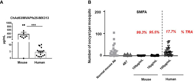
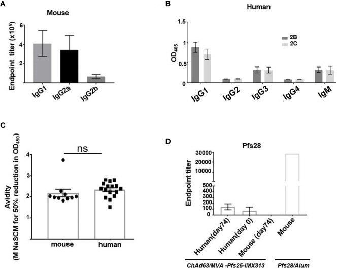
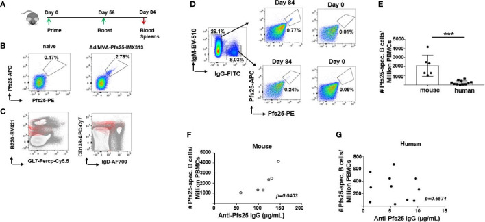
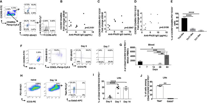
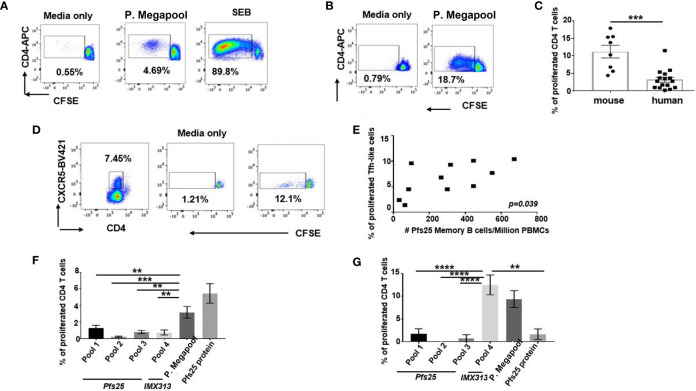
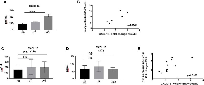
Plasmodium falciparum transmission-blocking vaccines (TBVs) targeting the Pfs25 antigen have shown promise in mice but the same efficacy has never been achieved in humans. We have previously published pre-clinical data related to a TBV candidate Pfs25-IMX313 encoded in viral vectors which was very promising and hence progressed to human clinical trials. The results from the clinical trial of this vaccine were very modest. Here we unravel why, contrary to mice, this vaccine has failed to induce robust antibody (Ab) titres in humans to elicit transmission-blocking activity. We examined Pfs25-specific B cell and T follicular helper (Tfh) cell responses in mice and humans after vaccination with Pfs25-IMX313 encoded by replication-deficient chimpanzee adenovirus serotype 63 (ChAd63) and the attenuated orthopoxvirus modified vaccinia virus Ankara (MVA) delivered in the heterologous prime-boost regimen via intramuscular route. We found that after vaccination, the Pfs25-IMX313 was immunologically suboptimal in humans compared to mice in terms of serum Ab production and antigen-specific B, CD4+ and Tfh cell responses. We identified that the key determinant for the poor anti-Pfs25 Ab formation in humans was the lack of CD4+ T cell recognition of Pfs25-IMX313 derived peptide epitopes. This is supported by correlations established between the ratio of proliferated antigen-specific CD4+/Tfh-like T cells, CXCL13 sera levels, and the corresponding numbers of circulating Pfs25-specific memory B cells, that consequently reflected on antigen-specific IgG sera levels. These correlations can inform the design of next-generation Pfs25-based vaccines for robust and durable blocking of malaria transmission.

Keywords: CD4+; P. falciparum; Pfs25; transmission blocking; vaccine.


Conflict of interest statement

AH is named inventor on patent applications covering malaria vaccines and immunization regimens. The remaining authors declare that the research was conducted in the absence of any commercial or financial relationships that could be construed as a potential conflict of interest.

Figures

Figure 1
Figure 1
Murine vaccine-induced antibodies demonstrate superior transmission-reducing activity to human. (A) Anti-Pfs25 serum total IgG responses for murine and human samples, as assessed on d72 post ChAd63/MVA Pfs25-IMX313 vaccination. Statistical analysis using 2-tailed Mann-Whitney test ***p < 0.001. (B) Transmission-reducing efficacy of human and murine anti-Pfs25 IgG induced by the ChAd63/MVA Pfs25-IMX313. Total Pfs25-specific IgG was purified from either pooled murine or individual human serum obtained on d72 post vaccination. The purified Pfs25-specific human and murine IgG samples were mixed with P. falciparum NF54 cultured gametocytes, at 100µg/ml and fed to (A) stephensi mosquitoes (n = 20 per test group). Murine samples were also tested at a lower concentration of 10µg/ml. IgG from naive mice was used as a negative control (“normal mouse Ab”); the transmission blocking anti-Pfs25 mAb 4β7 was used as a positive control. Midguts were dissected 8 days post-feeding. Transmission reducing activity (%TRA) for human and murine test IgG samples was calculated relative to the negative control IgG tested in the same assay and is indicated in red above each corresponding sample. Vertical dashed line separates human from murine samples. Data are from one out of three independent experiments giving similar results.
Figure 2
Figure 2
Assessment of murine and human antibodies’ effector functions, avidity and cross-reactivity. (A) Isotype profiles of murine Pfs25-specific antibody responses on d84 were assessed by ELISA. Responses are shown for IgG1, IgG2a and IgG2b. (B) Isotype profiles of human serum antibody responses were assessed by ELISA. Responses are shown for immunization groups 2B and 2C, assessed at d84 for IgG1, IgG2, IgG3, IgG4 and IgM. (C) Avidities of murine and human serum IgG responses at day 84 were assessed by NaSCN-displacement Pfs25 ELISA and are reported as the molar (M) concentration of NaSCN required to reduce the starting OD in the ELISA by 50% (IC50) Differences in avidity were assessed by Mann-Whitney test. Each symbol represents an individual response (ns- not significant). (D) Endpoint anti-Pfs28 IgG titers were assessed by ELISA on human serum samples isolated on d0 (n = 16) and d74 (n = 16) or murine samples (n = 6) obtained on d74 post ChAd63/MVA Pfs25-IMX313 vaccination. Murine serum sample acquired 2 weeks post boost immunization with recombinant Pfs28 protein in Alum, was used as positive control. Error bars show SEM.
Figure 3
Figure 3
Differences in murine and human Pfs25-specific B cell responses. (A) Experimental design in mice. (B) Representative FACS plots showing frequencies of Pfs25-specific B cells accessed among PBMCs isolated from naïve or ChAd63/MVA Pfs25-IMX313 vaccinated mice on d84. (C) Flow cytometry contour plots of B220 versus GL7 (left panel) or CD138 versus IgD (right panel) expression on PBMC suspensions of mice immunized with ChAd63/MVA Pfs25-IMX313 (gray contours). Pfs25-specific B220+ cells are represented as an overlay (red contours) to signify that the majority of antigen-specific B cells were indeed B220+,GL7-,CD138+,IgDlow. (D) Representative FACS plots showing frequencies of Pfs25-specific B cells among total IgM- or IgG-expressing memory B cells from PBMCs of trial vaccinees isolated on d0 and d84 post ChAd63/MVA Pfs25-IMX313 vaccination. (E) Bar graphs summarizing total numbers of Pfs25-specific B cells detected among individual murine and human PBMCs on d84 post vaccination. Data represent the mean ± SEM. Mann-Whitney test 2-tailed was performed to compare the two groups: ***p < 0.001. Correlations between serum anti-Pfs25 IgG concentrations and corresponding numbers of circulating antigen-specific B cells assessed for murine (F) and human (G) samples. P values were calculated using nonparametric Spearman’s test and are shown in the graphs.
Figure 4
Figure 4
Differences in murine and human Tfh cell responses. (A) Flow cytometric plots of the frequency of ICOS+PD1+ cells among either CD4+CD3+CXCR5+CD45RA+ or CD4+CD3+CXCR5+CD45RA- cells in the peripheral blood of healthy UK volunteers at d63 after ChAd63/MVA Pfs25-IMX313 vaccination. Flow cytometric assessment of the expression of CXCR3 and CCR6 within ICOS+PD1+CD4+CD3+CXCR5+CD45RA- cells is also shown. Correlations are shown between serum anti-Pfs25 IgG concentrations measured on d72 post vaccination and corresponding changes in numbers of either circulating (B) CD4+CD3+CXCR5+CD45RA- cells or (C) ICOS+PD1+CD4+CD3+CXCR5+CD45RA- cells on d28 relative to d0, or corresponding changes in numbers of (D) ICOS+PD1+CD4+CD3+CXCR5+CD45RA+ cells on d63 relative to d0. P values were calculated using nonparametric Spearman’s test and are shown in graphs. (E) Bars summarising frequencies of CXCR3-CCR6- (Th2-like), CXCR3+CCR6-(Th1-like) and CXCR3- CCR6+ (Th17-like) Tfh cell populations, among ICOS+PD1+CD4+CD3+CXCR5+CD45RA- cells as analyzed by flow cytometry. ****P < 0.0001, by one-way ANOVA, error bars show SEM. (F) Representative flow cytometric plots of the frequency of ICOS+PD1+ cells among CD4+CD3+CXCR5+CD44+CD62L- cells in the peripheral blood of vaccinated mice assessed on d0 and d7 post vaccination. (G) Total numbers of ICOS+PD1+CD4+CD3+CXCR5+CD44+CD62L- cells detected in peripheral blood of vaccinated mice at indicated time points. (H) Flow cytometry analyses of ICOS+PD1+CD4+CD3+CXCR5+CD44+CD62L- cells isolated from popliteal LNs of either naïve (d0) or vaccinated mice at d7 post ChAd63 Pfs25-IMX313 immunization. Assessment of intracellular Tbet and Gata3 expression on those cells is also shown (right panel). Summarised data are shown in (I) and (J). **P < 0.01, ****P < 0.0001, by Wilcoxon matched-pairs test, error bars show SEM.
Figure 5
Figure 5
Assessment of antigen specificity of murine and human Tfh cells. Tfh cells were quantified among human PBMCs (n = 14) or murine splenocytes (n = 6) isolated on d14 after vaccination with ChAd63/MVA Pfs25-IMX313.Antigen-specific, among total Tfh cells were identified either by OX40 upregulation in combination with ICOS+ (A) or CD25 co-expression (B) or CD154 expression (C) following 18 hours of stimulation with media only or megapool of 20-mer synthetic peptide sets encompassing the Pfs-25-IMX313 insert (P. megapool). Error bars show SEM. ns - not significant, P > 0.05, *P < 0.05, **P < 0.01, ***P < 0.001, ****P < 0.0001, by Wilcoxon matched-pairs test.
Figure 6
Figure 6
Proliferative capacity of vaccine-elicited murine and human CD4+ T cells. Cryopreserved and thawed human PBMCs (A) or murine splenocytes (B) isolated on d63 post vaccine administration were labelled with CFSE, stimulated with either media only or pool of stimulatory 20-mer peptides covering the entire Pfs25-IMX313 insert (P.megapool) for 6 days and analysed using flow cytometry for dividing (decreased CFSE) CD4+ T cells. As a positive control some cells were stimulated with staphylococcal enterotoxin B (SEB). Summarized data is shown in (C) Mann-Whitney test 2-tailed was performed to compare the two groups: ***p < 0.001. (D) Flow cytometric assessment of dividing human CXCR5+CD4+ cells in response to stimulation with media only or P. megapool is shown. (E) Correlation between total numbers of Pfs25-specific memory B cells (CD19+CD20+CD27+) detected on d84 among PBMCs of vaccinated healthy adult individuals and the percentage of their corresponding CD4+ T cells that divided CFSE in a response to P.megapool stimulation is shown. P value was calculated using nonparametric Spearman’s test and is shown in the graph. Percentage of human (F) and murine (G) CD4+ T cells that divided CFSE in a response to stimulation with either P. megapool, recombinant Pfs25 protein or separate stimulation with peptide pools 1-4,where pools 1-3 cover Pfs25 and pool 4 covers IMX313 portion of the vaccine construct. **P < 0.01, ***P < 0.001, ****P < 0.0001, by one-way ANOVA.
Figure 7
Figure 7
(A) CXCL13 was measured in murine serum following immunization at indicated time points by ELISA. Data represent mean ± SEM. Significance was calculated using ANOVA, ***P ≤ 0.001. (B) Correlation of change in CXCL13 levels on d63 relative to baseline with percentage of CD4+ T cells able to divide CFSE in a response to P.megapool stimulation. Correlation analysis performed using nonparametric Spearman’s test with 2-tailed P value, which is shown in the graph. CXCL13 was measured in serum of ChAd63/MVA Pfs25-IMX313 vaccinated trial individuals enrolled to groups 2B (C) or 2C (D) at indicated time points by ELISA. Data represent mean ± SEM. Significance was calculated using one-way ANOVA, ns-P > 0.05. (E) Correlation between changes in CXCL13 levels and numbers of ICOS+PD1+CD4+CD3+CXCR5+CD45RA+ cells both assessed on d63 post vaccination relative to baseline is shown. Correlation analysis performed using nonparametric Spearman’s test with 2-tailed P value, which is shown in the graph.

Similar articles

Cited by

References

    1. Thompson EA, Ols S, Miura K, Rausch K, Narum DL, Spangberg M, et al. . TLR-Adjuvanted Nanoparticle Vaccines Differentially Influence the Quality and Longevity of Responses to Malaria Antigen Pfs25. JCI Insight (2018) 3(10):e120692. doi: 10.1172/jci.insight.120692 - DOI - PMC - PubMed
    1. Acquah FK, Adjah J, Williamson KC, Amoaha LE. Transmission-Blocking Vaccines: Old Friends and New Prospects. Infect Immun (2019) 87(6):e00775–18. doi: 10.1128/IAI.00775-18 - DOI - PMC - PubMed
    1. Hoffman SL, Vekemans J, Richie TL, Duffy PE. The March Toward Malaria Vaccines. Vaccine (2015) 33 Suppl 4(Suppl 4):D13–23. doi: 10.1016/j.vaccine.2015.07.091 - DOI - PMC - PubMed
    1. Wang RB, Smith JD, Kappe SHI. Advances and Challenges in Malaria Vaccine Development. Expert Rev Mol Med (2009) 11:e39. doi: 10.1017/S1462399409001318 - DOI - PMC - PubMed
    1. Miura K, Deng B, Tullo G, Diouf A, Moretz SE, Locke E, et al. . Qualification of Standard Membrane-Feeding Assay With Plasmodium Falciparum Malaria and Potential Improvements for Future Assays. PloS One (2013) 8(3):e57909. doi: 10.1371/journal.pone.0057909 - DOI - PMC - PubMed

Publication types

MeSH terms