. 2021 Sep 29:12:757350.
doi: 10.3389/fgene.2021.757350. eCollection 2021.

Transcriptome Profiling Reveals Important Transcription Factors and Biological Processes in Skin Regeneration Mediated by Mechanical Stretch

Affiliations

Transcriptome Profiling Reveals Important Transcription Factors and Biological Processes in Skin Regeneration Mediated by Mechanical Stretch

Wei Liu et al. Front Genet. .

Abstract

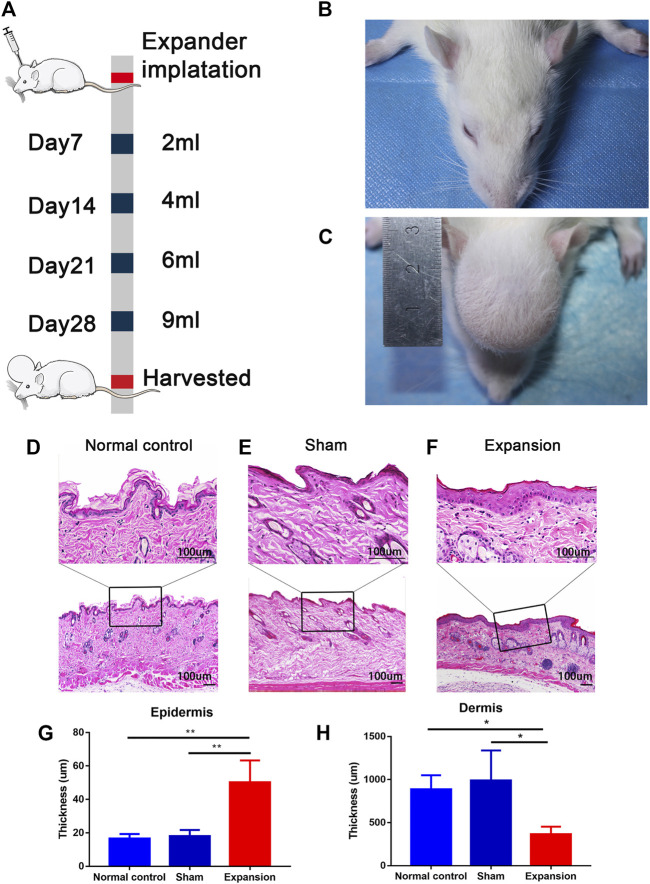
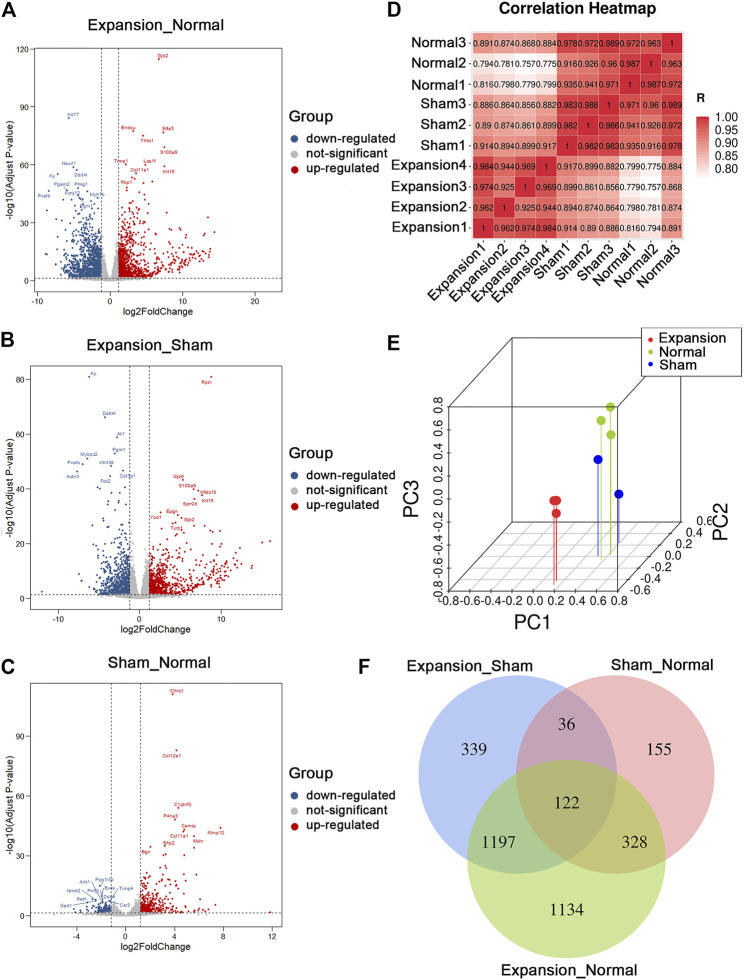
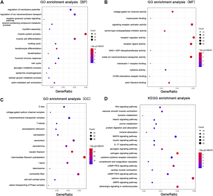
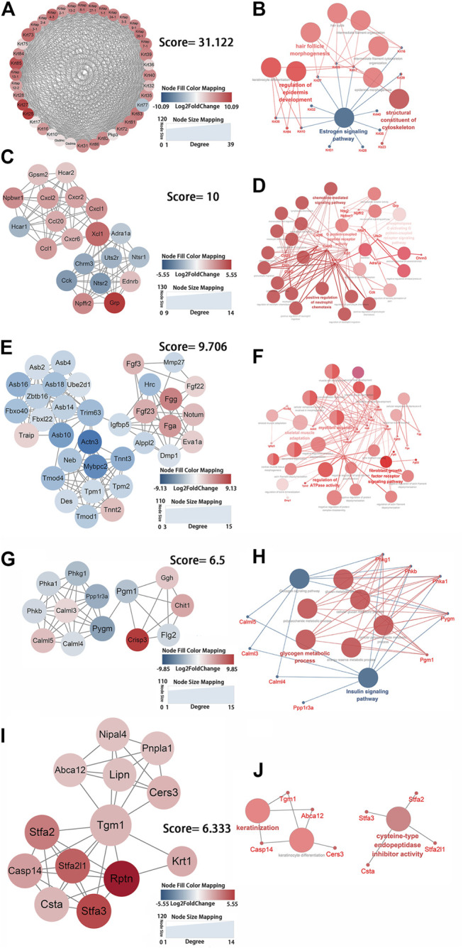
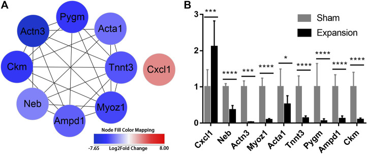
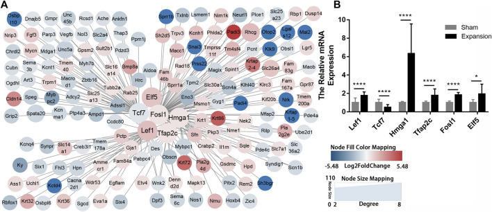
Background: Mechanical stretch is utilized to promote skin regeneration during tissue expansion for reconstructive surgery. Although mechanical stretch induces characteristic morphological changes in the skin, the biological processes and molecular mechanisms involved in mechanically induced skin regeneration are not well elucidated. Methods: A male rat scalp expansion model was established and the important biological processes related to mechanical stretch-induced skin regeneration were identified using Gene Ontology (GO) analysis, Kyoto Encyclopedia of Genes and Genomes (KEGG) analysis, and gene set enrichment analysis (GSEA). Analysis was also conducted by constructing a protein-protein interaction (PPI) network, identifying key modules and hub genes, determining transcription factor (TF)-mRNA regulatory relationships, and confirming the expression pattern of the TFs and hub genes. Results: We identified nine robust hub genes (CXCL1, NEB, ACTN3, MYOZ1, ACTA1, TNNT3, PYGM, AMPD1, and CKM) that may serve as key molecules in skin growth. These genes were determined to be involved in several important biological processes, including keratinocyte differentiation, cytoskeleton reorganization, chemokine signaling pathway, glycogen metabolism, and voltage-gated ion channel activity. The potentially significant pathways, including the glucagon signaling pathway, the Wnt signaling pathway, and cytokine-cytokine receptor interaction, were distinguished. In addition, we identified six TFs (LEF1, TCF7, HMGA1, TFAP2C, FOSL1, and ELF5) and constructed regulatory TF-mRNA interaction networks. Conclusion: This study generated a comprehensive overview of the gene networks underlying mechanically induced skin regeneration. The functions of these key genes and the pathways in which they participate may reveal new aspects of skin regeneration under mechanical strain. Furthermore, the identified TF regulators can be used as potential candidates for clinical therapeutics for skin pretreatment before reconstructive surgery.

Keywords: hub genes; mechanical stretch; skin regeneration; tissue expansion; transcription facotrs; transcriptome.


Conflict of interest statement

The authors declare that the research was conducted in the absence of any commercial or financial relationships that could be construed as a potential conflict of interest.

Figures

FIGURE 1
FIGURE 1
Overview of the study design and histological observation corresponding with morphological change of skin induced by mechanical stretch. (A) Schematic presentation of procedural timeline and expansion volume of scalp skin expansion. (B–C) Representative image of samples located on top of the dome induced by the skin expander and the central region of scalp in the sham operated and normal control group. (D–H) H and E staining showed a thicker epidermis and a thinner dermis thickness in the expanded group (n = 4) than that in the normal control group (n = 3) and the sham operated group (n = 3). scale bar: 100 um *p < 0.05, **p < 0.01.
FIGURE 2
FIGURE 2
Summary of differentially expressed genes and presentation of comprehensive similarity among the expanded group, the sham operated group, and the normal control group. (A) Analysis of differentially expressed genes between the expanded group and the normal control group by volcano mapping. (B) Analysis of differentially expressed genes between the expanded group and the sham operated group by volcano mapping. (C) Analysis of differentially expressed genes between the sham operated group and the normal control group by volcano mapping. (D) HeatMap of correlation analysis with unsupervised hierarchical clustering among the expanded, sham-operated, and normal control group. (E) 3D scatter of PCA among the expanded, sham operated, and normal control group. (F) Venn diagram showed the overlap of differentially expressed genes induced by mechanical stretch (blue + green, DEGs between the sham and normal groups were removed from those between the expanded and normal groups. The remaining DEGs were compared with those between the expanded and sham groups to obtain overlapping 1197 DEGs).
FIGURE 3
FIGURE 3
GO enrichment analysis and KEGG analysis showed changes in the molecular response and signaling pathway changes of skin during tissue expansion. (A) GO enrichment analysis of the DEGs in biological processes. (B) GO enrichment analysis of the DEGs in molecular functions. (C) GO enrichment analysis of the DEGs in cellular components. (D) KEGG analysis of the DEGs. Gene ratio indicates the number of DEGs associated with the GO term divided by the total number of DEGs. The size of the dots represents the number of DEGs associated with the GO term and the color represents the negative value of log10 of adjusted p-value. BP biological processes, MF molecular functions, CC cellular components.
FIGURE 4
FIGURE 4
Identification of the key protein clusters using string database and MCODE plugin (nodes >10, score >5) and functional analysis of identified 1-5 key protein clusters using ClueGo plugin. (A) Color coded network of protein cluster 1 and their connection. (B) Bubble plot of functional enrichment analysis showing proteins in cluster 1 participated in a structural constituent of keratin filament and intermediate filament cytoskeleton. (C) Color coded network of protein cluster 2 and their connection. (D) Bubble plot of functional enrichment analysis showing proteins in cluster 2 participated in chemokine signaling pathway. (E) Color coded network of protein cluster 3 and their connection. (F) Bubble plot of functional enrichment analysis showing proteins in cluster 3 participated in myofibril assembly. (G) Color coded network of protein cluster 4 and their connection. (H) Bubble plot of functional enrichment analysis showing proteins in cluster 4 participated in glycogen metabolic process (I) Color coded network of protein cluster 5 and their connection. (J) Bubble plot of functional enrichment analysis showing proteins in cluster 5 participated in keratinocyte differentiation. The nodes in (A) (C) (E) (G) (I) indicate proteins. The edges represent protein interaction. The bubbles in (B) (D) (F) (H) (J) indicate GO term, the dots represent proteins, and the edges represent the proteins enriched in GO term.
FIGURE 5
FIGURE 5
Identification of hub genes activated by mechanical stretch and their expression confirmed by qPCR (A) Hub genes identified with PPI analysis (B) The relative mRNA levels of CXCL1, NEB, ACTN3, MYOZ1, ACTA1, TNNT3, PYGM, AMPD1, and CKM using qPCR. *p < 0.05, ***p < 0.001, ****p < 0.0001.
FIGURE 6
FIGURE 6
Construction of regulatory networks of TF-mRNA interactions using transcription factor databases (A) Regulatory networks of TF-mRNA interactions activated by mechanical stretch (B) The relative mRNA levels of LEF1, TCF7, ELF5, FOSL1, TFAP2C, and HMGA1 using qPCR. *p < 0.05, ****p < 0.0001.

Similar articles

Cited by

References

    1. Adam R. C., Yang H., Ge Y., Lien W. H., Wang P., Zhao Y., et al. (2018). Temporal Layering of Signaling Effectors Drives Chromatin Remodeling during Hair Follicle Stem Cell Lineage Progression. Cell stem cell 22, 398–e7. 10.1016/j.stem.2017.12.004 - DOI - PMC - PubMed
    1. Addison C. L., Daniel T. O., Burdick M. D., Liu H., Ehlert J. E., Xue Y. Y., et al. (2000). The CXC Chemokine Receptor 2, CXCR2, Is the Putative Receptor for ELR+CXC Chemokine-Induced Angiogenic Activity. J. Immunol. 165, 5269–5277. 10.4049/jimmunol.165.9.5269 - DOI - PubMed
    1. Aragona M., Sifrim A., Malfait M., Song Y., Van Herck J., Dekoninck S., et al. (2020). Mechanisms of Stretch-Mediated Skin Expansion at Single-Cell Resolution. Nature 584, 268–273. 10.1038/s41586-020-2555-7 - DOI - PMC - PubMed
    1. Arcidiacono B., Chiefari E., Messineo S., Bilotta F. L., Pastore I., Corigliano D. M., et al. (2018). HMGA1 Is a Novel Transcriptional Regulator of the FoxO1 Gene. Endocrine 60, 56–64. 10.1007/s12020-017-1445-8 - DOI - PMC - PubMed
    1. Argyle D., Kitamura T. (2018). Targeting Macrophage-Recruiting Chemokines as a Novel Therapeutic Strategy to Prevent the Progression of Solid Tumors. Front. Immunol. 9, 2629. 10.3389/fimmu.2018.02629 - DOI - PMC - PubMed