. 2021 Oct 5:2021:2556352.
doi: 10.1155/2021/2556352. eCollection 2021.

Traditional Chinese Medicine Yang-Gan-Wan Alleviated Experimental Hepatic Damage by Inhibiting Oxidation, Inflammation, and Apoptosis in Cell and Mouse Models

Affiliations

Traditional Chinese Medicine Yang-Gan-Wan Alleviated Experimental Hepatic Damage by Inhibiting Oxidation, Inflammation, and Apoptosis in Cell and Mouse Models

Chia-Wen Yeh et al. Evid Based Complement Alternat Med. .

Abstract

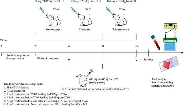
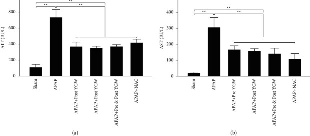
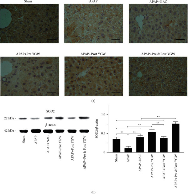
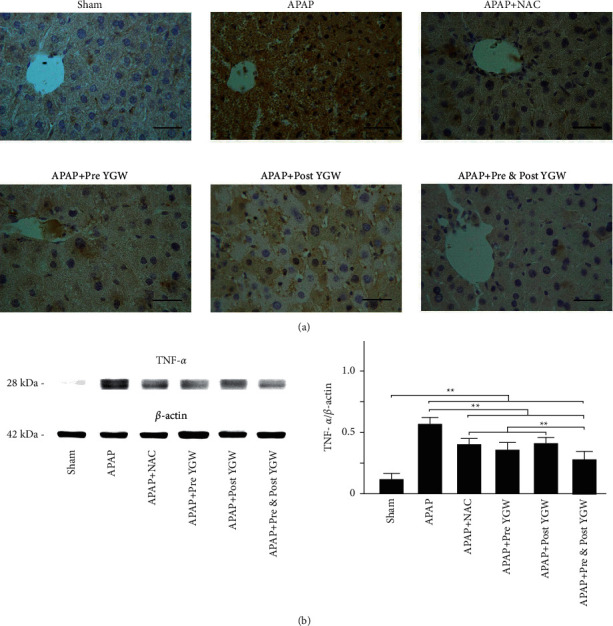
A hepatoprotective medicine, Yang-Gan-Wan (YGW), was used to treat hepatic damage in cell and mouse models. We performed a 1,1-diphenyl-2- picrylhydrazyl (DPPH) assay and found that YGW exhibited a significantly high free radical scavenging ability. Furthermore, the results of the 3-(4,5-dimethylthiazol-2-yl)-2,5-diphenyltetrazolium bromide (MTT) assay revealed that YGW treatment could alleviate lipopolysaccharide (LPS)-induced damage in Kupffer cells (liver macrophages). Enzyme-linked immunosorbent assay results demonstrated that YGW treatment could alleviate LPS-induced inflammation in Kupffer cells by inhibiting the expression of tumor necrosis factor (TNF)-α and interleukin (IL)-1β. By quantifying the serum levels of alanine aminotransferase (ALT) and aspartate aminotransferase (AST), we found that YGW treatment could alleviate hepatic damage and improve immunity in acetaminophen- (APAP-) treated mice by inhibiting the expression of ALT and AST. The findings of hematoxylin and eosin and Masson's trichrome staining indicated that YGW treatment could alleviate hepatic damage and reduce collagen fiber formation in the liver tissue of APAP-treated mice. Furthermore, immunohistochemistry staining and Western blot results showed that YGW treatment could alleviate oxidative stress, inflammation, and apoptosis in the liver tissue of APAP-treated mice by enhancing superoxide dismutase 2 (SOD2) expression but inhibiting TNF-α and caspase 3 expression. Our results suggest that YGW treatment exerted hepatoprotective effects on LPS-treated Kupffer cells and APAP-treated mice by inhibiting oxidation, inflammation, and apoptosis.


Conflict of interest statement

The authors declare that there are no conflicts of interest.

Figures

Figure 1
Figure 1
3D chromatographic fingerprint analysis of the hepatoprotective medicine YGW by using 3D HPLC. The following bioactive marker substances were identified: albiflorin and paeoniflorin from Paeonia lactiflora, prim-O-glucosylcimifugin and 5-O-methylvisammioside from Saposhnikovia divaricate, acteoside from Plantago asiatica, and ferulic acid and ligustilide from Angelica sinensis and Ligusticum striatum. AU, arbitrary perfusion units.
Figure 2
Figure 2
Antioxidative capacity of YGW. (a) DPPH assay using various concentrations of YGW treatment for 30 minutes. L-ascorbic acid is a standard antioxidative compound. (b) The DPPH free radical scavenging activity of YGW significantly increased with their concentration (n = 3 for each group; values are presented as mean ± SEM, ∗∗P < 0.01, P < 0.05, one-way ANOVA followed by the Student–Newman–Keuls multiple comparison post hoc test).
Figure 3
Figure 3
YGW treatments effectively alleviated LPS-induced damage in Kupffer cells. (a) Damaged and elongated Kupffer cells (indicated by the arrow) were observed after treatment with 1 μg/mL LPS; their morphology was restored to normal after treatment with 15 mg/mL YGW for 24 and 72 h scale bar = 30 μm. (b) MTT assay showed the cell viability of liver Kupffer cells under different concentrations of YGW. Irrespective of LPS treatment, the viability of Kupffer cells treated with YGW (5–20 mg/mL) significantly increased compared with that of Kupffer cells not treated with YGW (n = 3 for each group; values are presented as mean ± SEM).
Figure 4
Figure 4
YGW treatment effectively alleviated LPS-induced inflammation in Kupffer cells for 24 hours. The expression of the inflammatory markers TNF-α (a) and IL-1β (b) in Kupffer cells was significantly increased after LPS treatment but significantly decreased after YGW treatment (n = 3 for each group; values are presented as mean ± SEM).
Figure 5
Figure 5
Experimental design that draws the timeline of the action of YGW, LPS, and APAP, and the dosage of drugs and the time points of analyses. The dosage of YGW, APAP, and NAC was also shown. IP: intraperitoneal injection; OG: oral gavage.
Figure 6
Figure 6
YGW treatment effectively alleviated APAP-induced liver injury in mice. The expression of the liver injury-related markers (a) ALT and (b) AST significantly increased after APAP treatment but significantly decreased in the blood of mice after separating pre-YGW, post-YGW, pre- and post-YGW, and NAC treatments. N-Acetylcysteine (NAC) was the positive control for APAP treatment (n = 3 for each group; values are presented as mean ± SEM).
Figure 7
Figure 7
Oral YGW treatment effectively alleviated hepatic damage and reduced collagen fiber formation in APAP-treated mice. (a) H&E staining (200x and 400x) revealed normal liver cells in mice given the sham and YGW treatments but damaged liver cells in mice treated with APAP. After NAC, pre-YGW, post-YGW, and pre- and post-YGW treatment, the damaged liver tissue showed varying degrees of repair. (b) Masson's trichrome staining (200x and 400x) showed collagen fiber formation in the liver tissue of mice treated with APAP (blue color), whereas collagen fiber formation was partly restored in the liver tissue of APAP-treated mice after NAC, pre-YGW, post-YGW, and pre- and post-YGW treatments.
Figure 8
Figure 8
Oral YGW treatment effectively alleviated oxidative stress in the liver tissue of APAP-treated mice. (a) IHC staining showed antioxidant-related SOD2 expression in the liver tissue of sham-treated mice and APAP-treated mice after NAC, pre-YGW, post-YGW, and pre- and post-YGW treatments. Scale bar = 30 μm. (b) SOD2 expression was significantly lower in the liver tissue of mice treated with APAP compared with that in APAP-treated mice after NAC, pre-YGW, post-YGW, and pre- and post-YGW treatments (n = 3 for each group; values are presented as mean ± SEM).
Figure 9
Figure 9
Oral YGW treatment effectively alleviated inflammation in the liver tissue of APAP-treated mice. (a) IHC staining showed the expression of the inflammatory marker TNF-α in the liver tissue of sham-treated mice and APAP-treated mice after NAC, pre-YGW, post-YGW, and pre- and post-YGW treatment. Scale bar = 30 μm. (b) TNF-α expression was significantly higher in the liver tissue of APAP-treated mice compared with that in APAP-treated mice after NAC, pre-YGW, post-YGW, and pre- and post-YGW treatments (n = 3 for each group; values are presented as mean ± SEM).
Figure 10
Figure 10
Oral YGW treatment effectively alleviated apoptosis in the liver tissue of APAP-treated mice. (a) IHC staining showed the expression of the apoptosis-related marker c-caspase 3 in the liver tissue of sham-treated mice and APAP-treated mice after NAC, pre-YGW, post-YGW, and pre- and post-YGW treatments. Scale bar = 30 μm. (b) c-caspase 3/caspase 3 expression was significantly higher in the liver tissue of APAP-treated mice than in that of APAP-treated mice after NAC, pre-YGW, post-YGW, and pre- and post-YGW treatments (n = 3 for each group; values are presented as mean ± SEM).
Figure 11
Figure 11
Schematic illustrating that YGW treatment could alleviate liver injury in APAP-treated mice by inhibiting oxidative stress, inflammation, and apoptosis.

References

    1. Joung J. Y., Cho J. H., Kim Y. H., Choi S. H., Son C. G. A literature review for the mechanisms of stress‐induced liver injury. Brain and Behavior . 2019;9(3) doi: 10.1002/brb3.1235.e01235 - DOI - PMC - PubMed
    1. Zhang H. Y., Wang H. L., Zhong G. Y., Zhu J. X. Molecular mechanism and research progress on pharmacology of traditional Chinese medicine in liver injury. Pharmaceutical Biology . 2018;56(1):594–611. doi: 10.1080/13880209.2018.1517185. - DOI - PMC - PubMed
    1. Sakaida I., Matsumura Y., Akiyama S., Hayashi K., Ishige A., Okita K. Herbal medicine Sho-saiko-to (TJ-9) prevent liver fibrosis and enzyme-altered lesions in rat liver cirrhosis induced by a choline-deficient l-amino acid-defined diet. Journal of Hepatology . 1998;28(2):298–306. doi: 10.1016/0168-8278(88)80017-5. - DOI - PubMed
    1. Shimizu I., Ma Y.-R., Mizobuchi Y., et al. Effects of Sho-saiko-to, a Japanese herbal medicine, on hepatic fibrosis in rats. Hepatology . 1999;29(1):149–160. doi: 10.1002/hep.510290108. - DOI - PubMed
    1. Huang Y.-T., Lee T.-Y., Lin H.-C., Chou T.-Y., Yang Y.-Y., Hong C.-Y. Hemodynamic effects of Salvia miltiorrhiza on cirrhotic rats. Canadian Journal of Physiology and Pharmacology . 2001;79(7):566–572. doi: 10.1139/y01-029. - DOI - PubMed