. 2021 Dec;22(6):1360.
doi: 10.3892/etm.2021.10795. Epub 2021 Sep 24.

Long non-coding RNA NEAT1 contributes to lipopolysaccharide-induced inflammation and apoptosis of human middle ear epithelial cells via regulating the miR-301b-3p/TLR4 axis

Affiliations

Long non-coding RNA NEAT1 contributes to lipopolysaccharide-induced inflammation and apoptosis of human middle ear epithelial cells via regulating the miR-301b-3p/TLR4 axis

Zhuohui Liu et al. Exp Ther Med. 2021 Dec.

Abstract

Acute otitis media (AOM) is a common infectious disease in children that is accompanied by signs and symptoms of middle ear inflammation and infection. Previous studies have shown that the long non-coding (lnc)RNA nuclear-enriched abundant transcript 1(NEAT1) participates in various inflammatory conditions and plays an important regulatory role. The focus of the present study was the biological function of NEAT1 and underlying molecular mechanism in lipopolysaccharide (LPS)-induced human middle ear epithelial cells (HMEECs). The expression of NEAT1, miR-301b-3p and toll-like receptor 4 (TLR4) protein were determined by reverse transcription-quantitative PCR and western blot assays, respectively. Dual-luciferase reporter assay was performed to investigate the combination of miR-301b-3p and NEAT1 or TLR4. In addition, cell viability, apoptosis and the levels of pro-inflammatory factors (IL-1β, TNF-α and IL-6) were measured by Cell Counting Kit-8 assay, flow cytometry and ELISA, respectively. Cell viability was significantly decreased, whereas apoptosis and inflammation were increased in LPS-stimulated HMEECs. Functional analyses demonstrated that NEAT1 was upregulated following LPS treatment, whereas knockdown of NEAT1 significantly increased cell viability and alleviated apoptosis and inflammation. Mechanistically, NEAT1 directly bound to and negatively regulated miR-301b-3p expression, whereas miR-301b-3p inhibitors abolished the inhibitory effect of NEAT1 knockdown on cell apoptosis and inflammation. As a target of miR-301b-3p, TLR4 was regulated by NEAT1 and miR-301b-3p. TLR4 overexpression alleviated NEAT1 silencing-induced inflammatory suppression. Rescue experiments demonstrated that NEAT1 promoted TLR4 expression by inhibiting miR-301b-3p. Collectively, the results of the present study suggested that NEAT1 may attenuate LPS-induced inflammation and apoptosis in HMEECs by modulating the miR-301b-3p/TLR4 axis, and may provide a new therapeutic target for the clinical treatment of AOM.

Keywords: acute otitis media; human middle ear epithelial cells; miR-301b-3p; nuclear-enriched abundant transcript 1; toll-like receptor 4.


Conflict of interest statement

The authors declare that they have no competing interests.

Figures

Figure 1
Figure 1
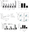
LPS reduces cell viability, and increases apoptosis and inflammatory response in human middle ear epithelial cells. (A and B) Cell viability was estimated by Cell Counting Kit-8 assay. *P<0.05, **P<0.01 vs. 0 µg. (C) Cell apoptosis was evaluated by flow cytometry. **P<0.01 vs. control. (D) IL-6, IL-1β and TNF-α expression was evaluated by ELISA. ***P<0.001 vs. control. LPS, lipopolysaccharide; OD, optical density; FITC, fluorescein isothi-ocyanate; PI, propidium iodide.
Figure 2
Figure 2
NEAT1 knockdown alleviates cell apoptosis and inflammatory response in LPS-induced human middle ear epithelial cells. (A) The expression of different long non-coding RNAs and (B) transfection efficiency of si-NEAT1 were analyzed by reverse transcription-quantitative PCR. (C) Cell viability was analyzed by Cell Counting Kit-8 assay. (D) Cell apoptosis was evaluated by flow cytometry. (E) IL-6, IL-1β and TNF-α expression was measured by ELISA. **P<0.01 and ***P<0.001 vs. control; #P<0.05, ##P<0.01 and ###P<0.001 vs. LPS+si-NC. LPS, LPS-stimulated; NEAT1, nuclear enriched abundant transcript 1; TUG1, taurine upregulated gene 1; ZFAS1, zinc finger antisense 1; PVT1, plasmacytoma variant translocation 1; ZEB1-AS1, zinc finger E-box binding homeobox 1 antisense 1; XIST, X-inactive-specific transcript; LPS, lipopolysaccharide; NC, negative control; si, small interfering; OD, optical density; FITC, fluorescein isothiocyanate; PI, propidium iodide.
Figure 3
Figure 3
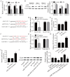
miR-301b-3p directly targets both NEAT1 and TLR4. (A) miR-301b-3p expression was detected by RT-qPCR. (B) TLR4 protein level was analyzed with western blotting. (C) Binding site of miR-301b-3p and NEAT1. (D) Dual-luciferase reporter assay was used to prove NEAT1 can target miR-301b-3p. (E) NEAT1 silencing significantly increased miR-301b-3p expression in HMEECs. (F) Binding site of miR-301b-3p and TLR4. (G) Dual-luciferase reporter assay was used to prove that miR-301b-3p can target TLR4. (H) NEAT1 expression was determined by RT-qPCR. (I) miR-301b-3p expression was detected by RT-qPCR. (J) TLR4 protein expression was measured by western blotting. *P<0.05 vs. control or NC-mimics; **P<0.01 vs. control, NC-mimics or si-NC; and ***P<0.001 vs. control or NC-mimics; ##P<0.01 compared with pc-vector. HMEECs, human middle ear epithelial cells; NEAT1, nuclear enriched abundant transcript 1; TLR4, toll-like receptor 4; LPS, lipopolysaccharide; miR, microRNA; NC, negative control; si, small interfering; WT, wild-type; MUT, mutant; RT-qPCR, reverse transcription-quantitative PCR.
Figure 4
Figure 4
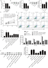
NEAT1 regulates LPS-induced cell viability, apoptosis and inflammation of human middle ear epithelial cells via the miR-370-3p/TLR4 axis. (A) miR-301b-3p expression was determined by reverse transcription-quantitative PCR. TLR4 protein expression was detected by western blot assay after transfection with (B) si-TLR4 and (C) pc-TLR4. (D) Cell viability was analyzed by Cell Counting Kit-8 assay. (E) Cell apoptosis was evaluated by flow cytometry. (F) IL-6, IL-1β and TNF-α expression levels were detected by ELISA. (G) TLR4 protein expression in different groups was detected by western blot assay. **P<0.01 vs. control, si-NC or pc-NC; ##P<0.01 vs. control and LPS+si-NC; &P<0.05 vs. LPS+si-NEAT1; &&P<0.01 vs. LPS+si-NEAT1; $$P<0.01 vs. LPS+si-TLR4. LPS, lipopolysaccharide; miR, microRNA; NC, negative control; si, small interfering; NEAT1, nuclear enriched abundant transcript 1; TLR4, toll-like receptor 4; OD, optical density; FITC, fluorescein isothiocyanate; PI, propidium iodide.

Similar articles

Cited by

References

    1. Morris LM, DeGagne JM, Kempton JB, Hausman F, Trune DR. Mouse middle ear ion homeostasis channels and intercellular junctions. PLoS One. 2012;7(e39004) doi: 10.1371/journal.pone.0039004. - DOI - PMC - PubMed
    1. Block SL. Causative pathogens, antibiotic resistance and therapeutic considerations in acute otitis media. Pediatr Infect Dis J. 1997;16:449–456. doi: 10.1097/00006454-199704000-00029. - DOI - PubMed
    1. Post JC. Direct evidence of bacterial biofilms in otitis media 2001. Laryngoscope. 2015;125:2003–2014. doi: 10.1002/lary.25291. - DOI - PubMed
    1. Topcuoglu N, Keskin F, Ciftci S, Paltura C, Kulekci M, Ustek D, Kulekci G. Relationship between oral anaerobic bacteria and otitis media with effusion. Int J Med Sci. 2012;9:256–261. doi: 10.7150/ijms.4382. - DOI - PMC - PubMed
    1. Beermann J, Piccoli MT, Viereck J, Thum T. Non-coding RNAs in development and disease: Background, mechanisms, and therapeutic approaches. Physiol Rev. 2016;96:1297–1325. doi: 10.1152/physrev.00041.2015. - DOI - PubMed