. 2021 Oct 7:2021:9925647.
doi: 10.1155/2021/9925647. eCollection 2021.

The DNA Repair Enzyme XPD Is Partially Regulated by PI3K/AKT Signaling in the Context of Bupivacaine-Mediated Neuronal DNA Damage

Affiliations

The DNA Repair Enzyme XPD Is Partially Regulated by PI3K/AKT Signaling in the Context of Bupivacaine-Mediated Neuronal DNA Damage

Wei Zhao et al. Oxid Med Cell Longev. .

Abstract

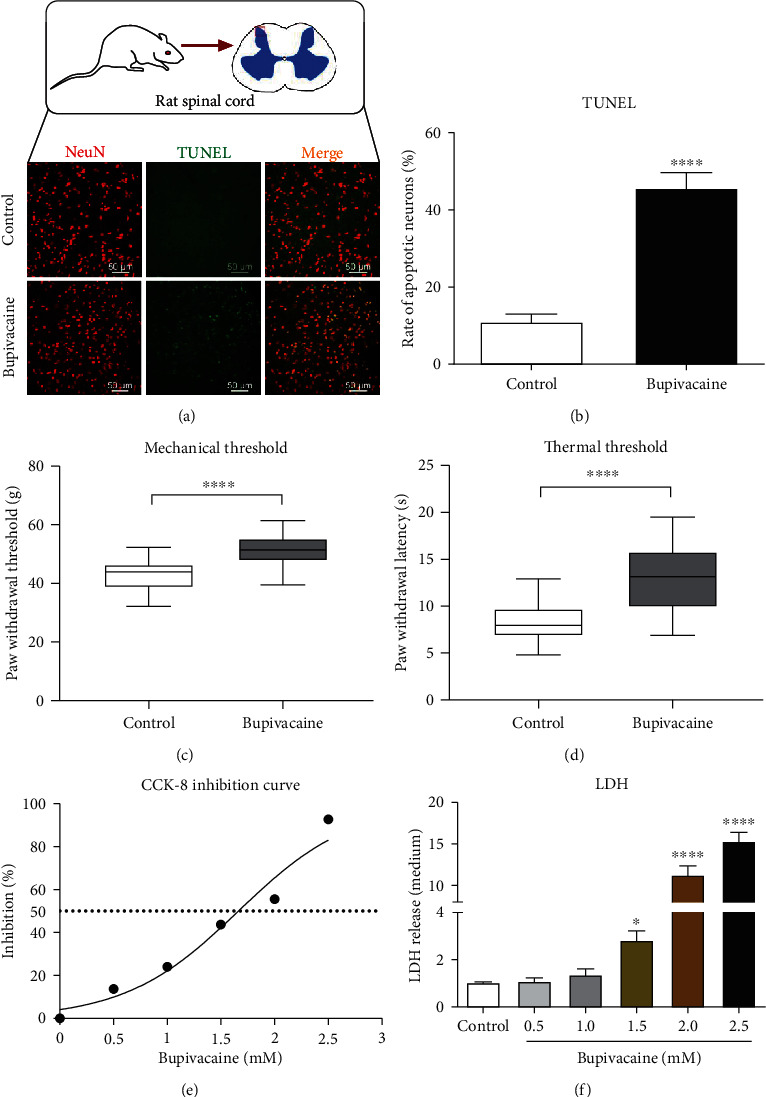
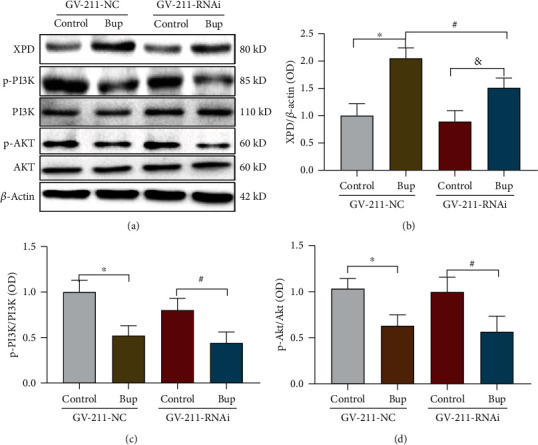
Bupivacaine, a local anesthetic widely used for regional anesthesia and pain management, has been reported to induce neuronal injury, especially DNA damage. Neurons employ different pathways to repair DNA damage. However, the mechanism underlying bupivacaine-mediated DNA damage repair is unclear. A rat neuronal injury model was established by intrathecal injection of (3%) bupivacaine. An in vitro neuronal injury model was generated by exposing SH-SY5Y cells to bupivacaine (1.5 mmol/L). Then, a cDNA plate array was used to identify the DNA repair genes after bupivacaine exposure. The results showed that xeroderma pigmentosum complementary group D (XPD) of the nuclear excision repair (NER) pathway was closely associated with the repair of DNA damage induced by bupivacaine. Subsequently, Western blot assay and immunohistochemistry indicated that the expression of the repair enzyme XPD was upregulated after DNA damage. Downregulation of XPD expression by a lentivirus aggravated the DNA damage induced by bupivacaine. In addition, phosphatidyl-3-kinase (PI3K)/AKT signaling in neurons was inhibited after exposure to bupivacaine. After PI3K/AKT signaling was inhibited, bupivacaine-mediated DNA damage was further aggravated, and the expression of XPD was further upregulated. However, knockdown of XPD aggravated bupivacaine-mediated neuronal injury but did not affect PI3K/AKT signaling. In conclusion, the repair enzyme XPD, which was partially regulated by PI3K/AKT signaling, responded to bupivacaine-mediated neuronal DNA damage. These results can be used as a reference for the treatment of bupivacaine-induced neurotoxicity.


Conflict of interest statement

The authors declare no competing interests.

Figures

Figure 1
Figure 1
Bupivacaine causes neurotoxic damage. In vivo, bupivacaine-induced apoptotic damage in rat spinal cord tissue and behavioral changes. The rats in the neuronal damage model group were intrathecally administered 20 μL of 3% bupivacaine, while rats in the control group were administered normal saline. Twenty-four hours after treatment with bupivacaine, the TUNEL staining was performed to determine the cell apoptosis rate ((a, b) n = 6; ∗∗∗∗p = 0.0001) in spinal cord tissue slices. NeuN was used to label mature neurons. TUNEL/NeuN positive cells (a) represent apoptotic neurons. The PWMT and PWTL were increased in the bupivacaine-treated group compared with the control group ((c, d) n = 6; ∗∗∗∗p = 0.0001). The in vivo data are shown as the mean ± SD. Bupivacaine may induce SH-SY5Y cell neurotoxicity in vitro. After SH-SY5Y cells were exposed to different concentrations of bupivacaine, the dose inhibition curve was plotted ((e) n = 3), and the IC50 of bupivacaine was calculated as 1.5 mmol/L. The release of LDH was determined to assess cytotoxicity ((f) n = 3; p = 0.0394 and ∗∗∗∗p < 0.0001 vs. the control group). The data are shown as the mean ± SD.
Figure 2
Figure 2
The expression of DNA damage repair genes was determined with a cDNA plate array after SH-SY5Y cells were exposed to bupivacaine. As shown in (a), a ratio > 0 indicates that the expression of the indicated gene was upregulated in the bupivacaine (Bup) group compared with the control (C) group, and a ratio < 0 indicates that the expression of the gene was downregulated. A ratio greater than 1 or less than -1 indicated that there was a one order of magnitude difference. A ratio greater than 2 or less than -2 indicated that there were two orders of magnitude difference. A heatmap of the 21 DNA repair proteins is shown in (b). Each column represents a sample (control group: Con1~3; bupivacaine group: Bup1~3), and each row represents one of the DNA repair proteins. The cDNA levels of the DNA repair genes are indicated by the different colors, with the color changing from green to red with increasing expression.
Figure 3
Figure 3
The expression of the crucial NER pathway-associated repair enzyme XPD was significantly increased after exposure to bupivacaine in vivo and in vitro. The mRNA expression of XPD (a) was significantly increased in bupivacaine-treated cells compared with control cells at different time points, especially at 12 h ((a) n = 3; ∗∗∗p = 0.0035, ∗∗∗∗p < 0.0001), and the protein expression of XPD was also significantly increased at different time points, especially at 24 h ((b, c) n = 3; ∗∗∗p = 0.001 and ∗∗∗∗p < 0.0001 vs. the control group). The expression of XPD in spinal cord tissue was significantly increased in the rat spinal neurotoxicity model compared with the control group (Western blotting: (d, e) n = 6; ∗∗∗∗p < 0.0001; immunohistochemistry: (f, g) n = 6; ∗∗p = 0.0028). The in vivo data are presented as the mean ± SD.
Figure 4
Figure 4
Downregulation of XPD expression further aggravated the neurotoxicity caused by bupivacaine. After XPD expression was downregulated with the XPD-GV211-RNAi-expressing lentivirus, the SH-SY5Y cell apoptosis and DNA damage induced by bupivacaine were exacerbated. GV211-NC served as a control lentivirus. DNA damage was aggravated in bupivacaine-treated SH-SY5Y cells in which XPD expression was inhibited (Bup-GV211-RNAi) compared to the control group (Bup-GV211-NC), as the phosphorylation of γ-H2AX, a DNA damage marker, was significantly increased ((a, b) n = 3; &p = 0.0243), (p = 0.0013, and #p = 0.0243, vs. the control group). Furthermore, the expression of the apoptosis-related proteins Bcl-2 and Bax was reduced ((a, c) n = 3; &p = 0.0100) (p = 0.0008, and #p < 0.0001 vs. the control group), and the olive tail moment was significantly higher in the comet assay ((d, e) n = 3; &p = 0.0002) (p < 0.0001, and #p < 0.0001 vs. the control group). Moreover, suppression of XPD expression significantly increased the apoptosis ratio, as determined by flow cytometry ((f, g) n = 3; &p = 0.0308) (p < 0.0412, and #p < 0.0003 vs. the control group). The data are shown as the mean ± SD of three independent experiments.
Figure 5
Figure 5
Inhibition of PI3K/AKT aggravated SH-SY5Y injury caused by bupivacaine and further increase the expression of XPD. Treatment with LY294002, a PI3K/AKT inhibitor, alone significantly increased XPD expression in SH-SY5Y cells ((a, b) n = 3; p = 0.0005). Moreover, the expression of XPD was further increased in the bupivacaine and LY294002-treated group compared with the bupivacaine-treated group ((a, b) n = 3; &p = 0.0254) (#p = 0.0003 vs. the control group). LY294002 (10 μM) further inhibited the protein expression of p-PI3K ((c, d) n = 3; #p = 0.0316) (p = 0.0308 vs. the control group) and p-AKT ((c, e) n = 3; #p = 0.0346) (p = 0.0198 vs. the control group) after bupivacaine treatment. The expression of the apoptosis-related proteins Bax and Bcl-2 and p-γH2AX (markers of DNA damage) was increased in the 10 μM LY294002 and bupivacaine-treated group compared with the bupivacaine group ((f) #p = 0.0115; (g) n = 3; #p = 0.0360) ((f) p = 0.0011 vs. the control group) ((g) p = 0.0003 vs. the control group). The data are shown as the mean ± SD of three independent experiments.
Figure 6
Figure 6
Downregulation of XPD expression did not affect the PI3K/AKT pathway. In SH-SY5Y cells, bupivacaine increased the expression of XPD ((a, b) n = 3; p = 0.0009 and &p = 0.0208 vs. the control group). The expression of XPD was downregulated by the XPD-GV211-RNAi-expressing lentivirus, and GV211-NC served as a control lentivirus ((a, b) n = 3; #p = 0.0413). p-PI3K and p-AKT expression was inhibited by bupivacaine ((a, c) p = 0.0060 and #p = 0.0291; (a, d) p = 0.0345 and #p = 0.0242 vs. the control group). Nevertheless, the expression of p-PI3K ((a, c) n = 3; p = 0.8535) and p-AKT ((a, d) n = 3; p = 0.9396) was not further reduced in the group in which XPD expression was downregulated compared with the GV-211-NC-treated group. The data are shown as the mean ± SD of three independent experiments.
Figure 7
Figure 7
Graphical abstract. Bupivacaine may cause neuronal oxidative DNA damage. Damaged DNA activates a largely unknown repair mechanism. Our study showed that XPD is closely involved in repairing bupivacaine-induced oxidative DNA damage in neurons. Notably, XPD may be partially regulated by the PI3K/AKT pathway.

References

    1. Meng Q. T., Xia Z. Y., Liu J., Bayliss D. A., Chen X. Local anesthetic inhibits hyperpolarization-activated cationic currents. Molecular Pharmacology . 2011;79(5):866–873. doi: 10.1124/mol.110.070227. - DOI - PMC - PubMed
    1. Kato J., Konishi J., Yoshida H., et al. Cauda equina syndrome following combined spinal and epidural anesthesia: a case report. Canadian Journal of Anesthesia/Journal canadien d'anesthésie . 2011;58(7):638–641. doi: 10.1007/s12630-011-9505-7. - DOI - PubMed
    1. Xiang Y., Wang W., Jing S., Zhang Z., Wang D. Vocal fold paralysis and cauda equina syndrome following spinal-epidural anesthesia. Medicine . 2021;100(3, article e24374) doi: 10.1097/MD.0000000000024374. - DOI - PMC - PubMed
    1. Poprac P., Jomova K., Simunkova M., Kollar V., Rhodes C. J., Valko M. Targeting free radicals in oxidative stress-related human diseases. Trends in Pharmacological Sciences . 2017;38(7):592–607. doi: 10.1016/j.tips.2017.04.005. - DOI - PubMed
    1. Kunkel T. A. Celebrating DNA's repair crew. Cell . 2015;163(6):1301–1303. doi: 10.1016/j.cell.2015.11.028. - DOI - PubMed

Substances