

Current limitations to identify covid-19 using artificial intelligence with chest x-ray imaging (part ii). The shortcut learning problem
- PMID: 34660166
- PMCID: PMC8502237
- DOI: 10.1007/s12553-021-00609-8
Current limitations to identify covid-19 using artificial intelligence with chest x-ray imaging (part ii). The shortcut learning problem
Abstract
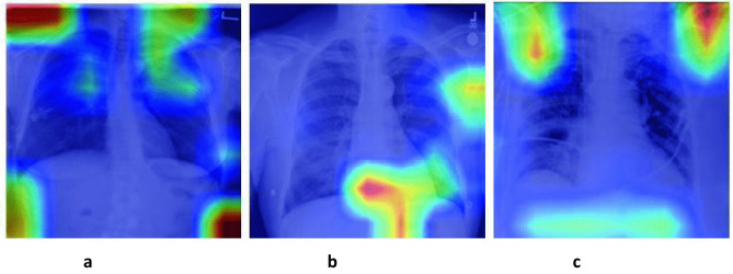
Since the outbreak of the COVID-19 pandemic, computer vision researchers have been working on automatic identification of this disease using radiological images. The results achieved by automatic classification methods far exceed those of human specialists, with sensitivity as high as 100% being reported. However, prestigious radiology societies have stated that the use of this type of imaging alone is not recommended as a diagnostic method. According to some experts the patterns presented in these images are unspecific and subtle, overlapping with other viral pneumonias. This report seeks to evaluate the analysis the robustness and generalizability of different approaches using artificial intelligence, deep learning and computer vision to identify COVID-19 using chest X-rays images. We also seek to alert researchers and reviewers to the issue of "shortcut learning". Recommendations are presented to identify whether COVID-19 automatic classification models are being affected by shortcut learning. Firstly, papers using explainable artificial intelligence methods are reviewed. The results of applying external validation sets are evaluated to determine the generalizability of these methods. Finally, studies that apply traditional computer vision methods to perform the same task are considered. It is evident that using the whole chest X-Ray image or the bounding box of the lungs, the image regions that contribute most to the classification appear outside of the lung region, something that is not likely possible. In addition, although the investigations that evaluated their models on data sets external to the training set, the effectiveness of these models decreased significantly, it may provide a more realistic representation as how the model will perform in the clinic. The results indicate that, so far, the existing models often involve shortcut learning, which makes their use less appropriate in the clinical setting.
Keywords: Artificial Intelligence; COVID-19; Chest X-Rays; Deep Learning.
© IUPESM and Springer-Verlag GmbH Germany, part of Springer Nature 2021.
Conflict of interest statement
Conflicts of interestThe authors declare that they have no conflict of interest.
Figures

Similar articles
-
Current limitations to identify COVID-19 using artificial intelligence with chest X-ray imaging.Health Technol (Berl). 2021;11(2):411-424. doi: 10.1007/s12553-021-00520-2. Epub 2021 Feb 5. Health Technol (Berl). 2021. PMID: 33585153 Free PMC article.
-
Prognostication of patients with COVID-19 using artificial intelligence based on chest x-rays and clinical data: a retrospective study.Lancet Digit Health. 2021 May;3(5):e286-e294. doi: 10.1016/S2589-7500(21)00039-X. Epub 2021 Mar 24. Lancet Digit Health. 2021. PMID: 33773969 Free PMC article.
-
COVID-19 Classification from Chest X-Ray Images: A Framework of Deep Explainable Artificial Intelligence.Comput Intell Neurosci. 2022 Jul 14;2022:4254631. doi: 10.1155/2022/4254631. eCollection 2022. Comput Intell Neurosci. 2022. PMID: 35845911 Free PMC article.
-
A narrative review on characterization of acute respiratory distress syndrome in COVID-19-infected lungs using artificial intelligence.Comput Biol Med. 2021 Mar;130:104210. doi: 10.1016/j.compbiomed.2021.104210. Epub 2021 Jan 18. Comput Biol Med. 2021. PMID: 33550068 Free PMC article. Review.
-
Biphasic majority voting-based comparative COVID-19 diagnosis using chest X-ray images.Expert Syst Appl. 2023 Apr 15;216:119430. doi: 10.1016/j.eswa.2022.119430. Epub 2022 Dec 21. Expert Syst Appl. 2023. PMID: 36570382 Free PMC article. Review.
Cited by
-
New patch-based strategy for COVID-19 automatic identification using chest x-ray images.Health Technol (Berl). 2022;12(6):1117-1132. doi: 10.1007/s12553-022-00704-4. Epub 2022 Nov 10. Health Technol (Berl). 2022. PMID: 36406188 Free PMC article.
-
Development and Validation of a Multimodal-Based Prognosis and Intervention Prediction Model for COVID-19 Patients in a Multicenter Cohort.Sensors (Basel). 2022 Jul 2;22(13):5007. doi: 10.3390/s22135007. Sensors (Basel). 2022. PMID: 35808502 Free PMC article.
-
Machine learning with multimodal data for COVID-19.Heliyon. 2023 Jul 5;9(7):e17934. doi: 10.1016/j.heliyon.2023.e17934. eCollection 2023 Jul. Heliyon. 2023. PMID: 37483733 Free PMC article. Review.
-
Implementation of artificial intelligence in thoracic imaging-a what, how, and why guide from the European Society of Thoracic Imaging (ESTI).Eur Radiol. 2023 Jul;33(7):5077-5086. doi: 10.1007/s00330-023-09409-2. Epub 2023 Feb 2. Eur Radiol. 2023. PMID: 36729173 Free PMC article. Review.
-
Improving deep neural network generalization and robustness to background bias via layer-wise relevance propagation optimization.Nat Commun. 2024 Jan 4;15(1):291. doi: 10.1038/s41467-023-44371-z. Nat Commun. 2024. PMID: 38177129 Free PMC article.
References
Publication types
LinkOut - more resources
Full Text Sources