. 2021 Oct 1:8:746837.
doi: 10.3389/fsurg.2021.746837. eCollection 2021.

Remote Training of Functional Endoscopic Sinus Surgery With Advanced Manufactured 3D Sinus Models and a Telemedicine System

Affiliations

Remote Training of Functional Endoscopic Sinus Surgery With Advanced Manufactured 3D Sinus Models and a Telemedicine System

Masanobu Suzuki et al. Front Surg. .

Abstract

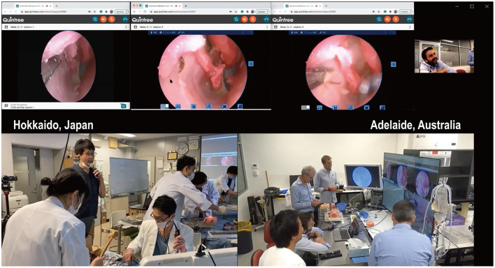
Objective: Traditionally, cadaveric courses have been an important tool in surgical education for Functional Endoscopic Sinus Surgery (FESS). The recent COVID-19 pandemic, however, has had a significant global impact on such courses due to its travel restrictions, social distancing regulations, and infection risk. Here, we report the world-first remote (Functional Endoscopic Sinus Surgery) FESS training course between Japan and Australia, utilizing novel 3D-printed sinus models. We examined the feasibility and educational effect of the course conducted entirely remotely with encrypted telemedicine software. Methods: Three otolaryngologists in Hokkaido, Japan, were trained to perform frontal sinus dissections on novel 3D sinus models of increasing difficulty, by two rhinologists located in Adelaide, South Australia. The advanced manufactured sinus models were 3D printed from the Computed tomography (CT) scans of patients with chronic rhinosinusitis. Using Zoom and the Quintree telemedicine platform, the surgeons in Adelaide first lectured the Japanese surgeons on the Building Block Concept for a three Dimensional understanding of the frontal recess. They in real time directly supervised the surgeons as they planned and then performed the frontal sinus dissections. The Japanese surgeons were asked to complete a questionnaire pertaining to their experience and the time taken to perform the frontal dissection was recorded. The course was streamed to over 200 otolaryngologists worldwide. Results: All dissectors completed five frontal sinusotomies. The time to identify the frontal sinus drainage pathway (FSDP) significantly reduced from 1,292 ± 672 to 321 ± 267 s (p = 0.02), despite an increase in the difficulty of the frontal recess anatomy. Image analysis revealed the volume of FSDP was improved (2.36 ± 0.00 to 9.70 ± 1.49 ml, p = 0.014). Questionnaires showed the course's general benefit was 95.47 ± 5.13 in dissectors and 89.24 ± 15.75 in audiences. Conclusion: The combination of telemedicine software, web-conferencing technology, standardized 3D sinus models, and expert supervision, provides excellent training outcomes for surgeons in circumstances when classical surgical workshops cannot be realized.

Keywords: 3D printer; building block concept; frontal sinusotomy; functional endoscopic sinus surgery (FESS); social distancing regulation; surgical training.


Conflict of interest statement

MS: receiving royalties from Igakushoin. PW: consultant for Fusetec, Neilmed and receiving royalties from Medtronic, Fusetec, and Integra. Shareholder for Chitogel. AP: consultant for Fusetec, Medtronic, Tissium, and ENT technologies. Shareholder for Chitogel and Speakers Bureau for Sequiris. The remaining authors declare that the research was conducted in the absence of any commercial or financial relationships that could be construed as a potential conflict of interest.

Figures

Figure 1
Figure 1
The remote endoscopic surgery training course was conducted between Hokkaido, Japan, and Adelaide, Australia, and broadcasted worldwide. The course was held over 2 days in February 2021. Three otolaryngologists in Hokkaido were trained to perform frontal sinus dissections on 3D sinus models printed from CT scans of patients. Two rhinologists based in Adelaide, 8,000-km away from Hokkaido, simultaneously viewed the surgeries and provided real-time feedback using a telemedicine platform, Quintree. The course was also broadcast worldwide to over 200 otolaryngologists' personal computers.
Figure 2
Figure 2
The remote endoscopic surgery training course was objectively and subjectively beneficial in improving the dissectors' surgical techniques. (A) The time to identify the FSDP. It was defined as the time interval from starting to resect the anterior wall of ANC to identify and insert a ball probe tip to FSDP at the height of the top of ANC. P-values for indicated comparisons were determined by t-test. *p < 0.05. (B) The relative time to identify the FSDP to the instructor's demonstration. P-values for indicated comparisons were determined by t-test. *p < 0.05. (C) The subjective difficulty of the surgeries that the dissectors were perceiving during the dissection. (D) The subjective completeness of frontal sinusotomy during the dissection. (E) The confidence for frontal sinusotomy in general before, during, and after the course. P-values for indicated comparisons were determined by t-test. *p < 0.05.
Figure 3
Figure 3
The volume of FSDP before and after the frontal sinusotomies and the frontal drill out. (A) 3D computed graphic images of right FSDP in the model 6 before and after the sinusotomies. (B) The volume of FSDP in 3D sinus models was significantly improved after the frontal sinusotomies than before the procedure. P-values for indicated comparisons were determined by t-test. *p < 0.05, **p < 0.01. (C) 3D computed graphic images of bilateral FSDP in the 3D sinus models before and after the frontal drillout (the model 2). (D) The total volume of bilateral FSDPs was also significantly improved after the frontal drillout. P-values for indicated comparisons were determined by t-test. *p < 0.05.
Figure 4
Figure 4
The result of questionnaire from the dissectors and the audiences on educational benefits of the remote FESS training course. A questionnaire survey was performed on dissectors and audiences, including the following topic; the general benefit of the course to improve the surgical skills (0: not at all to 100: extremely beneficial), the usefulness of instructions from Adelaide (0: not at all to 100: excellent), subjective distance from Adelaide (0: felt as if they were in Hokkaido to 100: far away), the dependency on the translation between English and Japanese (0: completely independent to 100: perfect dependent), the reproducibility of the 3D sinus models to the real nasal cavity and paranasal sinuses (0: not at all to 100: completely reproduced, for dissectors only), the reproducibility of the 3D sinus models to the real nasal cavity and paranasal sinuses when watched on monitors (0: not at all to 100: completely reproduced, for audiences only), the necessity of using cadavers for the similar surgical training (0: not at all to 100: essential), the quality of the communication by Quintree (0: terrible to 100: very good), and the educational effect of watching the three surgeries with same anatomies at the same time by Quintree (0: terrible to 100: very good). Data were gathered by visual-analog scale and expressed as the mean ± SD.

References

    1. Fokkens WJ, Lund VJ, Hopkins C, Hellings PW, Kern R, Reitsma S, et al. . European position paper on rhinosinusitis and nasal polyps 2020. Rhinology. (2020) 58(Suppl.S29):1–464. 10.4193/Rhin20.601 - DOI - PubMed
    1. Wormald P-J. Endoscopic Sinus Surgery. Stuttgart, New York, NY: Thieme (2018). 10.1055/b-0038-149997 - DOI
    1. Naidoo Y, Wen D, Bassiouni A, Keen M, Wormald PJ. Long-Term results after primary frontal sinus surgery. Int Forum Allergy Rhinol. (2012) 2:185–90. 10.1002/alr.21015 - DOI - PubMed
    1. Wormald PJ. Surgery of the frontal recess and frontal sinus. Rhinology. (2005) 43:82–5. - PubMed
    1. Wormald PJ. The agger nasi cell: the key to understanding the anatomy of the frontal recess. Otolaryngol Head Neck Surg. (2003) 129:497–507. 10.1016/S0194-5998(03)01581-X - DOI - PubMed

LinkOut - more resources