Review
. 2022 Feb;36(2):172-180.
doi: 10.1111/jdv.17744. Epub 2021 Nov 2.

Cutaneous findings following COVID-19 vaccination: review of world literature and own experience

Affiliations
Review

Cutaneous findings following COVID-19 vaccination: review of world literature and own experience

T Gambichler et al. J Eur Acad Dermatol Venereol. 2022 Feb.

Abstract

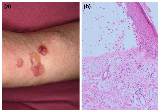
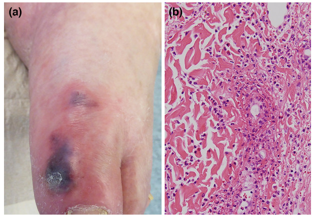
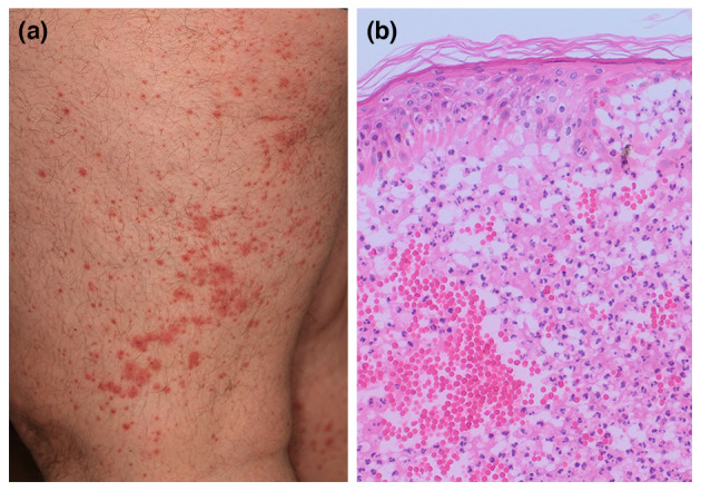
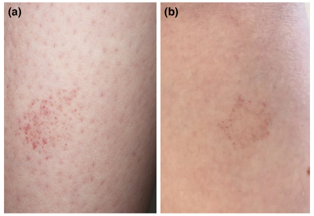
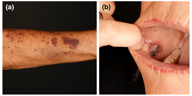
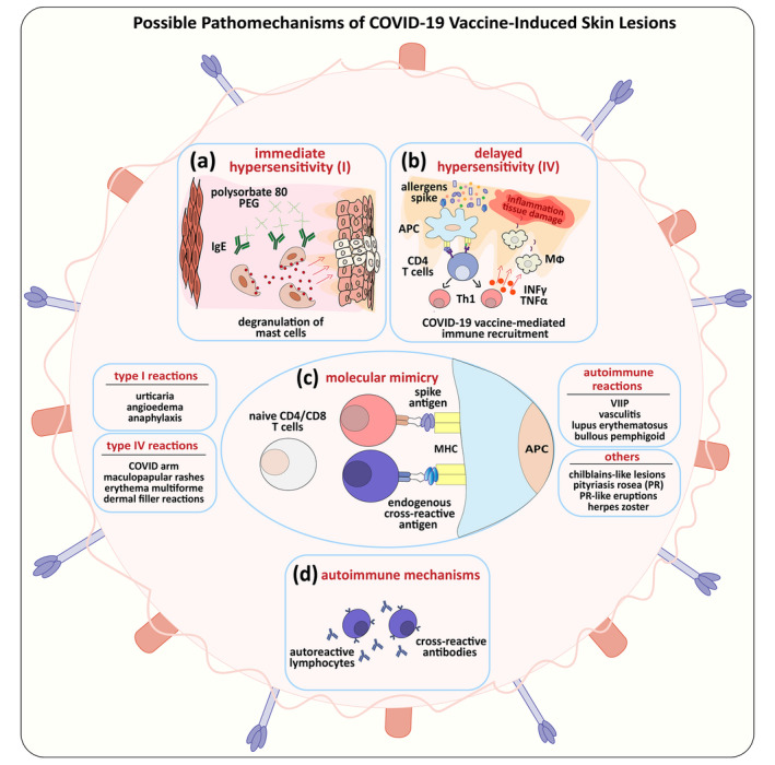
There is growing evidence that not only the novel coronavirus disease (COVID-19) but also the COVID-19 vaccines can cause a variety of skin reactions. In this review article, we provide a brief overview on cutaneous findings that have been observed since the emerging mass COVID-19 vaccination campaigns all over the world. Unspecific injection-site reactions very early occurring after the vaccination are most frequent. Type I hypersensitivity reactions (e.g. urticaria, angio-oedema and anaphylaxis) likely due to allergy to ingredients may rarely occur but can be severe. Type IV hypersensitivity reactions may be observed, including delayed large local skin lesions ("COVID arm"), inflammatory reactions in dermal filler or previous radiation sites or even old BCG scars, and more commonly morbilliform and erythema multiforme-like rashes. Autoimmune-mediated skin findings after COVID-19 vaccination include leucocytoclastic vasculitis, lupus erythematosus and immune thrombocytopenia. Functional angiopathies (chilblain-like lesions, erythromelalgia) may also be observed. Pityriasis rosea-like rashes and reactivation of herpes zoster have also been reported after COVID-19 vaccination. In conclusion, there are numerous cutaneous reaction patterns that may occur following COVID-19 vaccination, whereby many of these skin findings are of immunological/autoimmunological nature. Importantly, molecular mimicry exists between SARS-CoV-2 (e.g. the spike-protein sequences used to design the vaccines) and human components and may thus explain some COVID-19 pathologies as well as adverse skin reactions to COVID-19 vaccinations.


Figures

Figure 1
Figure 1
An old man had a history of intense itch and erythematous skin lesions after his first Comirnaty® vaccination. After the patients’ second Comirnaty® vaccination, erythematous‐bullous skin lesions (a) spread over his extremities and back. Serology, routine histopathology (b) as well as direct and indirect immunofluorescence confirmed the diagnosis of bullous pemphigoid that was most likely triggered by the vaccine. Under medium‐dose corticosteroids the itch and skin lesions gradually resolved.
Figure 2
Figure 2
Chilblain‐like lesions (a) on the left big toe of a patient who had received his first shot Comirnaty® about 2 weeks ago. One week ago, the same skin findings were noticed on his right big toe. Histopathology was consistent with a leucocytoclastic vasculitis (b). Complete work‐up (e.g. infection serologies, antibody tests, X‐ray, ultrasound) did not reveal relevant pathologies. Since there was no evidence for other causes, we made a diagnosis of vaccine‐induced leucocytoclastic vasculitis. The skin lesions gradually resolved after tapered systemic corticosteroids.
Figure 3
Figure 3
Widespread purpuric rash on the left thigh (a) of an elderly male which had developed a few days after his first Comirnaty® . He had no history for previous infections or new medication. Histopathology was consistent with a leucocytoclastic vasculitis (b). Complete work‐up (e.g. infection serologies, antibody tests, X‐ray, ultrasound) did not reveal relevant pathologies. A diagnose of vaccine‐induced leucocytoclastic vasculitis was made. The skin lesions gradually resolved after tapered systemic corticosteroids.
Figure 4
Figure 4
A young healthy female had a history of an asymptomatic petechial rash developing 1 day after her first Comirnaty® vaccination on her arms and legs. The discrete lesions spontaneously regressed within a few weeks. Six weeks later after her second Comirnaty® shot she presented with the same petechial macules, in part with bizarre annular configuration (a); histopathology revealed slight perivascular lymphocytic infiltrates, mild vacuolar changes with occasional necrotic keratinocytes and erythrocyte extravasates (b). On laboratory investigations, her thrombocytes and other coagulation parameters were within the normal range. Clinically and histopathologically, a vaccine‐induced Majocchi’s disease‐like rash was diagnosed. Again, her skin lesions slowly resolved without treatment after few weeks.
Figure 5
Figure 5
Showing a female patient with a 2‐week history of widespread dry purpuric skin lesions on her extremities (a). A few days prior to presentation in our hospital, she had a history for spontaneous gastrointestinal, genital, nasal and oral bleedings (b). Notably, about 2 weeks before the manifestation of purpuric skin lesions she had her first Vaxzevira ® shot. She was otherwise well‐being and had no new medications. On presentation, her thrombocytes counted 1000/µL (150.000–400.000/µL) and the immature thrombocyte fraction was 0.0% (1.1–6.1). A complete work‐up did not reveal potential causes for the thrombocytopenic purpura observed, except for her COVID‐19 vaccination. A diagnosis of vaccine‐induced immune thrombocytopenic purpura was made. Under high‐dose corticosteroids her thrombocytopenia gradually recovered.
Figure 6
Figure 6
Illustrating possible pathomechanisms of cutaneous findings following COVID‐19 vaccination. (a) Immediate (type I) allergic reactions to COVID19‐vaccine components, polyethylene glycols (PEG) and cross‐reactive polysorbate 80, can lead to mast cell degranulation causing urticaria, angio‐oedema and anaphylaxis. (b) In presence of secondary allergens, increased COVID‐19 vaccine‐mediated immune recruitment may manifest as delayed (type IV) allergic reactions, such as “COVID arm”, maculopapular rashes, erythema multiforme and dermal filler reactions. (c) Mechanism of molecular mimicry caused by genetic similarities of SARS‐COV‐2 spike protein components to endogenous cross‐reactive human antigens. (d) Generation of autoreactive lymphocytes and cross‐reactive antibodies due to molecular mimicry leading to autoimmune reactions, such as vaccine‐induced immune thrombocytopenia (VIIT) lupus erythematosus, vasculitis and bullous pemphigoid. Other COVID‐19 vaccine‐related observations include functional angiopathies such as chilblains‐like lesions and re‐activation of viral conditions such as pityriasis rosea (PR), PR‐like eruptions and herpes zoster [IgE = immunoglobulin E, APC = antigen‐presenting cells, INFγ = interferon γ, TNFα = tumour necrosis factor α, Th1 = type 1 T helper cells, MΦ = macrophages, MHC = major histocompatibility complex].

Comment in

References

    1. Kurup D, Schnell MJ. SARS‐CoV‐2 vaccines ‐ the biggest medical research project of the 21st century. Curr Opin Virol 2021; 49: 52–57. - PMC - PubMed
    1. Carneiro DC, Sousa JD, Monteiro‐Cunha JP. The COVID‐19 vaccine development: a pandemic paradigm. Virus Res 2021; 17: 198454. - PMC - PubMed
    1. Vasireddy D, Atluri P, Malayala SV, Vanaparthy R, Mohan G. Review of COVID‐19 Vaccines Approved in the United States of America for Emergency Use. J Clin Med Res 2021; 13: 204–213. - PMC - PubMed
    1. Li Y, Tenchov R, Smoot J, Liu C, Watkins S, Zhou Q. A comprehensive review of the global efforts on COVID‐19 vaccine development. ACS Central Sci 2021; 7: 512–533. - PMC - PubMed
    1. Blumental S, Debré P. Challenges and issues of anti‐SARS‐CoV‐2 vaccines. Front Med (Lausanne) 2021; 8: 664179. - PMC - PubMed

Substances