. 2021 Nov 23;15(11):17480-17498.
doi: 10.1021/acsnano.1c03905. Epub 2021 Oct 18.

Biomimetic Mechanically Strong One-Dimensional Hydroxyapatite/Poly(d,l-lactide) Composite Inducing Formation of Anisotropic Collagen Matrix

Affiliations

Biomimetic Mechanically Strong One-Dimensional Hydroxyapatite/Poly(d,l-lactide) Composite Inducing Formation of Anisotropic Collagen Matrix

Yonggang Zhang et al. ACS Nano. .

Abstract

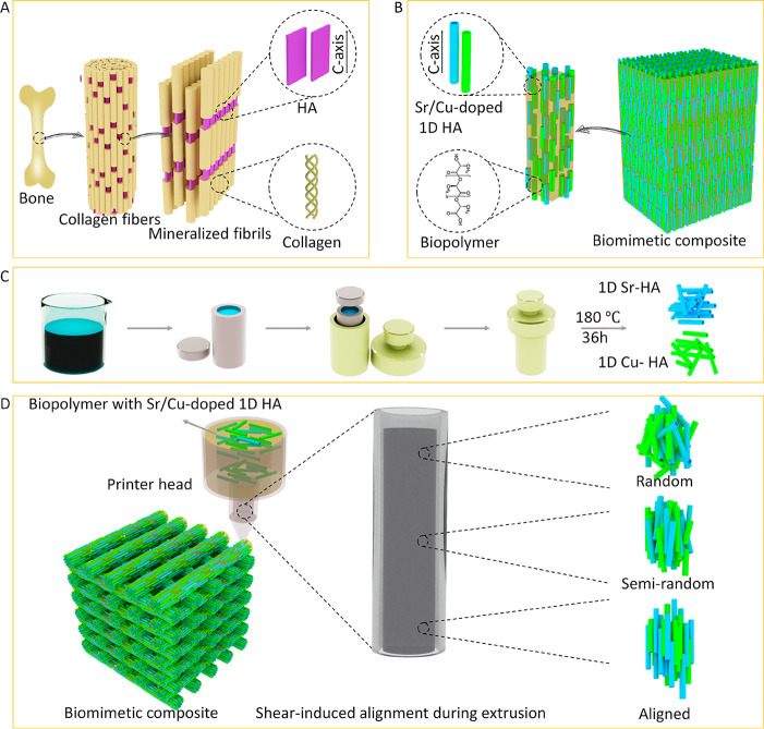
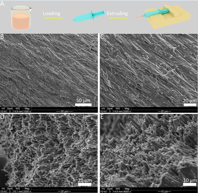
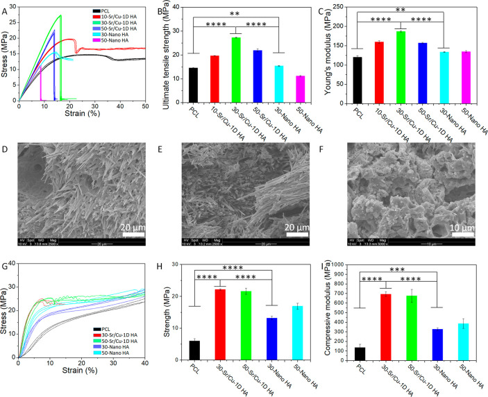
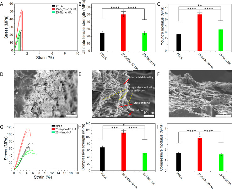
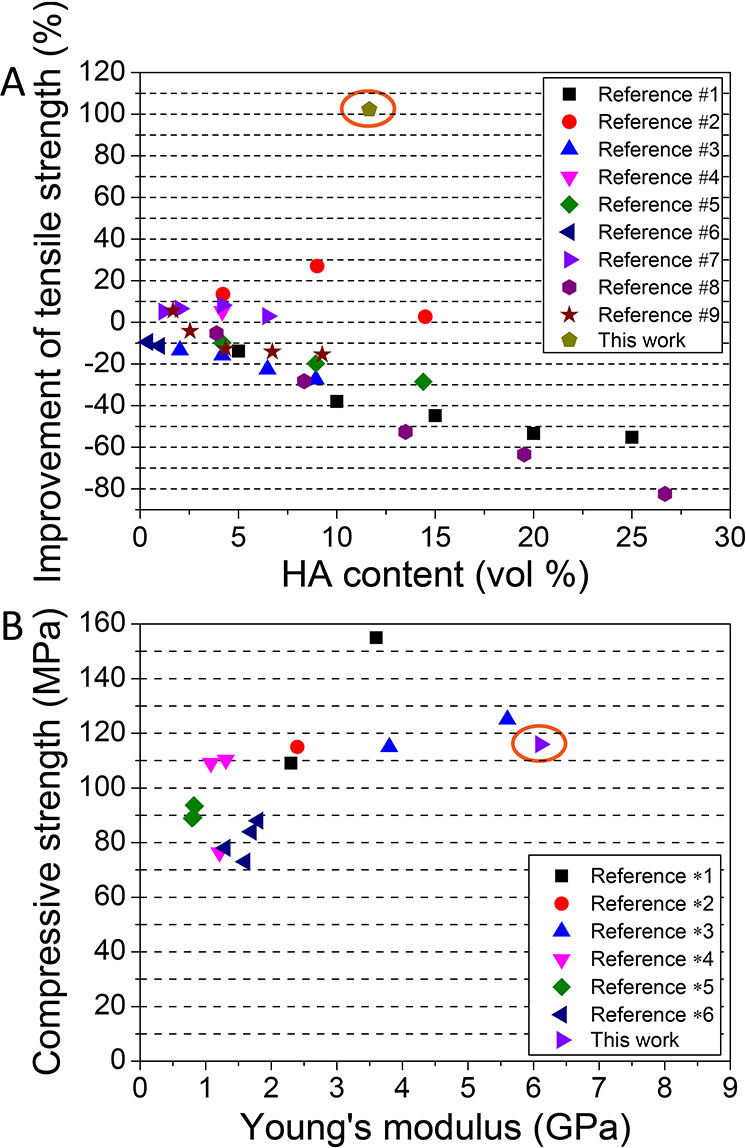
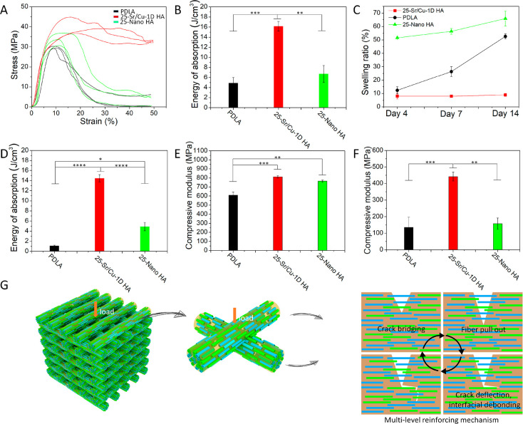
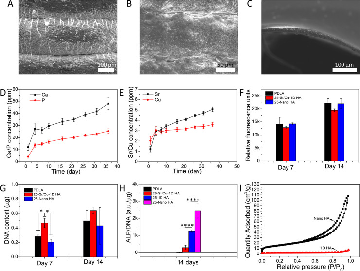
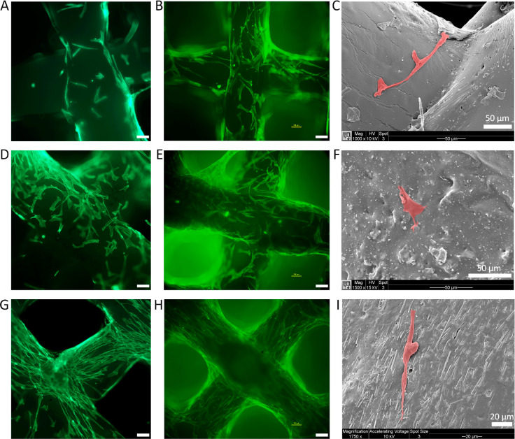
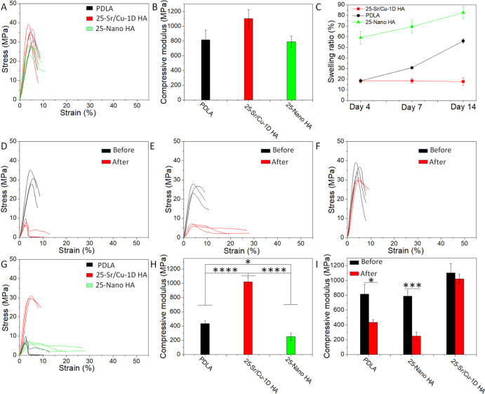
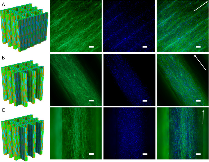
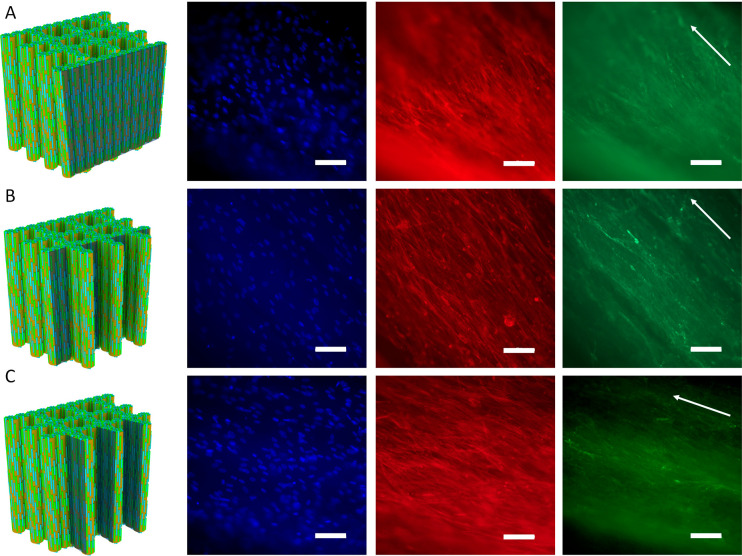
Natural bone is a complex composite, consisting predominantly of collagen and hydroxyapatite (HA), which form a highly organized, hierarchical structure from the nano- to the macroscale. Because of its biphasic, anisotropic, ultrafine structural design, bone tissue possesses excellent mechanical properties. Herein, inspired by the composition and microstructure of natural bone, a biphasic composite consisting of highly aligned strontium/copper-doped one-dimensional hydroxyapatite (Sr/Cu-doped 1D HA) and poly(d,l-lactide) (PDLA) was developed. The presence and alignment of Sr/Cu-doped 1D HA crystals resulted in mechanical reinforcement of the polymer matrix, including compressive and tensile strength and modulus, fracture toughness, swelling resistance, and long-term structural stability. The compressive strength, tensile strength, and Young's modulus of the biomimetic composite were comparable to that of cortical bone. Biologically, the biomimetic composite showed a sustained release of the incorporated Sr and Cu ions, facilitated mineral deposition from simulated body fluid, and supported attachment, proliferation, and alkaline phosphatase activity of human mesenchymal stromal cells (hMSCs). Moreover, the highly aligned Sr/Cu-doped 1D HA crystals in the 3D porous scaffolds induced the alignment of hMSCs and secretion of an anisotropic collagen fiber matrix in 3D. The biomimetic Sr/Cu-doped 1D HA/PDLA composite presented here contributes to the current efforts aiming at the design and development of load-bearing bioactive synthetic bone graft substitutes. Moreover, the biomimetic composite may serve as a 3D platform for studying cell-extracellular matrix interactions in bone tissue.

Keywords: PDLA; anisotropy; biomimetic; bone; composite; hydroxyapatite.


Conflict of interest statement

The authors declare no competing financial interest.

Figures

Figure 1
Figure 1
Schematic illustration of the (A) microstructure of natural bone, (B) design of the biomimetic composite, (C) preparation process of the 1D Sr-HA and 1D Cu-HA, and (D) fabrication process of the biomimetic composite.
Figure 2
Figure 2
(A) Schematic illustration of the fabrication process of Sr/Cu-doped 1D HA/PCL filaments by using a syringe. SEM micrographs of the surface (B, C) and cross-section (D, E) of the Sr/Cu-doped 1D HA/PCL filament obtained by extruding a suspension with the solid content of 33.3 wt % using an 18G needle with a length of 10 mm at the extrusion rate of 100 μL/min.
Figure 3
Figure 3
(A to C) SEM micrographs of Sr/Cu-doped 1D HA/PCL filaments obtained by extruding a suspension with the solid content of 33.3 wt % at the extrusion rate of 100 μL/min using needles with a length of 10 mm but different diameter (25G, 21G, 18G, respectively). (D to F) SEM micrographs of Sr/Cu-doped 1D HA/PCL filaments obtained by extruding a suspension with the solid content of 33.3 wt % at the extrusion rate of 100 μL/min using 21G needles with different lengths (2, 6, 10 mm, respectively). (G to I) SEM micrographs of Sr/Cu-doped 1D HA/PCL filaments obtained by extruding a suspension with the solid content of 33.3 wt % by using the same needle (21G, 2 mm) but three different extrusion rates (100, 200, 400 μL/min, respectively). (J to L) SEM micrographs of Sr/Cu-doped 1D HA/PCL filaments obtained by using the same needle (21G, 2 mm) and the same extrusion rate (100 μL/min) but three different concentrations of the solid content of the suspension (24.4, 33.3, 42.3 wt %, respectively).
Figure 4
Figure 4
(A–C) Tensile stress–strain curves, UTS, and Young’s modulus of pure PCL, biomimetic Sr/Cu-doped 1D HA/PCL composites with 10, 30, and 50 wt % Sr/Cu-doped 1D HA, and Nano HA/PCL composites with 30 and 50 wt % Nano HA, respectively. (D–F) SEM micrographs of the fracture surface of biomimetic Sr/Cu-doped 1D HA/PCL composites with 30 and 50 wt % Sr/Cu-doped 1D HA and the Nano HA/PCL composite with 50 wt % Nano HA after the tensile test, respectively. (G–I) Compressive stress–strain curves, compressive strength (at 5% strain), and compressive modulus of pure PCL, biomimetic Sr/Cu-doped 1D HA/PCL composites with 30 and 50 wt % Sr/Cu-doped 1D HA, and Nano HA/PCL composites with 30 and 50 wt % Nano HA in the longitudinal direction, respectively. (*) for p < 0.05, (**) for p < 0.01, (***) for p < 0.001, and (****) for p < 0.0001.
Figure 5
Figure 5
(A–C) Tensile stress–strain curves, UTS, and Young’s modulus of pure PDLA, the biomimetic Sr/Cu-doped 1D HA/PDLA composite with 25 wt % Sr/Cu-doped 1D HA, and the Nano HA/PDLA composite with 25 wt % Nano HA, respectively. (D–F) SEM micrographs of the fracture surface after the tensile test of pure PDLA, the biomimetic Sr/Cu-doped 1D HA/PDLA composite with 25 wt % Sr/Cu-doped 1D HA, and the Nano HA/PDLA composite with 25 wt % Nano HA, respectively. (G–I) Compressive stress–strain curves, compressive strength, and compressive modulus of pure PDLA, the biomimetic Sr/Cu-doped 1D HA/PDLA composite with 25 wt % Sr/Cu-doped 1D HA, and the Nano HA/PDLA composite with 25 wt % Nano HA, respectively. (*) for p < 0.05, (**) for p < 0.01, (***) for p < 0.001, and (****) for p < 0.0001.
Figure 6
Figure 6
Comparison of reinforcement efficiency of (A) tensile and (B) compressive strength of HA/PLA composites between this and previously published work (more information and references can be found in Table S5 and Table S6).
Figure 7
Figure 7
(A, B) Compressive stress–strain curves and energy of absorption per unit volume of the Sr/Cu-doped 1D HA/PDLA porous scaffolds, pure PDLA porous scaffolds, and Nano HA/PDLA porous scaffolds, respectively. (C) Swelling ratio of the Sr/Cu-doped 1D HA/PDLA porous scaffolds, pure PDLA porous scaffolds, and Nano HA/PDLA porous scaffolds in SBF at 37 °C for different time intervals (4, 7, and 14 days). (D) Energy of absorption per unit volume of the Sr/Cu-doped 1D HA/PDLA porous scaffolds, pure PDLA porous scaffolds, and Nano HA/PDLA porous scaffolds after a 14-day hMSC culture. (E, F) Compressive modulus of the pure PDLA porous scaffolds, Nano HA/PDLA porous scaffolds, and Sr/Cu-doped 1D HA/PDLA porous scaffolds before (E) and after (F) a 14-day hMSCs culture. (G) Schematic illustration of the proposed reinforcement mechanism of aligned Sr/Cu-doped 1D HA in the PDLA matrix. (*) for p < 0.05, (**) for p < 0.01, (***) for p < 0.001, and (****) for p < 0.0001.
Figure 8
Figure 8
(A–C) In vitro mineralization of Sr/Cu-doped 1D HA/PDLA porous scaffolds (A), Nano HA/PDLA porous scaffolds (B), and pure PDLA porous scaffolds (C) in SBF at 37 °C for 7 days. (D) Cumulative amounts of Ca and P in physiological saline solution in time upon immersion of Sr/Cu-doped 1D HA/PDLA porous scaffolds. (E) Cumulative amounts of Sr and Cu in physiological saline solution in time upon immersion of Sr/Cu-doped 1D HA/PDLA porous scaffolds. (F, G) Metabolic activity and DNA content of hMSCs cultured for 7 and 14 days on pure PDLA porous scaffolds, Nano HA/PDLA porous scaffolds, and Sr/Cu-doped 1D HA/PDLA porous scaffolds. (H) ALP activity of hMSCs cultured for 14 days on pure PDLA porous scaffolds, Sr/Cu-doped 1D HA/PDLA porous scaffolds, undoped 1D HA/PDLA porous scaffolds, and Nano HA/PDLA porous scaffolds, respectively. (I) N2 adsorption–desorption isotherms of Nano HA and undoped 1D HA. (*) for p < 0.05, (**) for p < 0.01, (***) for p < 0.001, and (****) for p < 0.0001.
Figure 9
Figure 9
(A, B) Fluorescence microscopy images of actin cytoskeleton (phalloidin: green) staining of hMSCs cultured on pure PDLA porous scaffolds for 3 and 7 days, respectively. (C) SEM micrographs of hMSCs cultured on pure PDLA porous scaffolds for 3 days. (D, E) Fluorescence microscopy images of actin cytoskeleton (phalloidin: green) staining of hMSCs cultured on Nano HA/PDLA porous scaffolds for 3 and 7 days, respectively. (F) SEM micrographs of hMSCs cultured on Nano HA/PDLA porous scaffolds for 3 days. (G, H) Fluorescence microscopy images of actin cytoskeleton (phalloidin: green) staining of hMSCs cultured on Sr/Cu-doped 1D HA/PDLA porous scaffolds for 3 and 7 days, respectively. (I) SEM micrographs of hMSCs cultured on Sr/Cu-doped 1D HA/PDLA porous scaffolds for 3 days. Unlabeled scale bar: 100 μm.
Figure 10
Figure 10
(A, B) Compressive stress–strain curves and compressive modulus of the assembled Sr/Cu-doped 1D HA/PDLA porous scaffolds, assembled PDLA porous scaffolds, and assembled Nano HA/PDLA porous scaffolds, respectively. (C) Swelling ratio of the assembled Sr/Cu-doped 1D HA/PDLA porous scaffolds, assembled PDLA porous scaffolds, and assembled Nano HA/PDLA porous scaffolds in SBF at 37 °C for different time intervals (4, 7, and 14 days). (D–F) Compressive stress–strain curves of the assembled PDLA porous scaffolds (D), assembled Nano HA/PDLA porous scaffolds (E), and assembled Sr/Cu-doped 1D HA/PDLA porous scaffolds (F) before and after a 14-day hMSC culture. (G, H) Compressive stress–strain curves and compressive modulus of the assembled Sr/Cu-doped 1D HA/PDLA porous scaffolds, assembled PDLA porous scaffolds, and assembled Nano HA/PDLA porous scaffolds after a 14-day hMSC culture. (I) Compressive modulus of the assembled PDLA porous scaffolds, assembled Nano HA/PDLA porous scaffolds, and assembled Sr/Cu-doped 1D HA/PDLA porous scaffolds before and after a 14-day hMSC culture. (*) for p < 0.05, (**) for p < 0.01, (***) for p < 0.001, and (****) for p < 0.0001.
Figure 11
Figure 11
(A–C) Fluorescence microscopy images of actin cytoskeleton (phalloidin: green) and nuclei (DAPI: blue) staining of hMSCs cultured on the assembled Sr/Cu-doped 1D HA/PDLA porous scaffolds for 4 days. Arrows indicate the direction of the ceramic alignment. Scale bar: 100 μm.
Figure 12
Figure 12
(A–C) Fluorescence microscopy images of nuclei (DAPI: blue), actin cytoskeleton (phalloidin: red), and type I collagen (green) of hMSCs cultured on the assembled Sr/Cu-doped 1D HA/PDLA porous scaffolds for 14 days. Arrows indicate the direction of the ceramic alignment. Scale bar: 100 μm.

References

    1. Bohner M.; Miron R. J. A Proposed Mechanism for Material-Induced Heterotopic Ossification. Mater. Today 2019, 22, 132–141. 10.1016/j.mattod.2018.10.036. - DOI
    1. Wu S. L.; Liu X. M.; Yeung K. W. K.; Liu C. S.; Yang X. J. Biomimetic Porous Scaffolds for Bone Tissue Engineering. Mater. Sci. Eng., R 2014, 80, 1–36. 10.1016/j.mser.2014.04.001. - DOI
    1. Woodruff M. A.; Lange C.; Reichert J.; Berner A.; Chen F.; Fratzl P.; Schantz J.-T.; Hutmacher D. W. Bone Tissue Engineering: From Bench to Bedside. Mater. Today 2012, 15 (10), 430–435. 10.1016/S1369-7021(12)70194-3. - DOI
    1. Christy P. N.; Basha S. K.; Kumari V. S.; Bashir A. K. H.; Maaza M.; Kaviyarasu K.; Arasu M. V.; Al-Dhabi N. A.; Ignacimuthu S. Biopolymeric Nanocomposite Scaffolds for Bone Tissue Engineering Applications – A Review. J. Drug Delivery Sci. Technol. 2020, 55, 101452.10.1016/j.jddst.2019.101452. - DOI
    1. Rezwan K.; Chen Q. Z.; Blaker J. J.; Boccaccini A. R. Biodegradable and Bioactive Porous Polymer/Inorganic Composite Scaffolds for Bone Tissue Engineering. Biomaterials 2006, 27 (18), 3413–3431. 10.1016/j.biomaterials.2006.01.039. - DOI - PubMed

Publication types