. 2021 Oct 18;13(1):192.
doi: 10.1186/s13148-021-01125-2.

SETD2-mediated epigenetic regulation of noncanonical Wnt5A during osteoclastogenesis

Affiliations

SETD2-mediated epigenetic regulation of noncanonical Wnt5A during osteoclastogenesis

Moonmoon Deb et al. Clin Epigenetics. .

Abstract

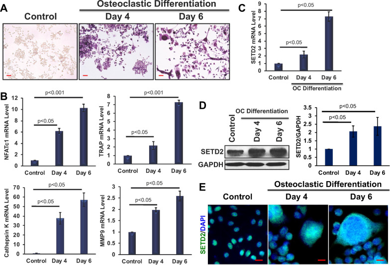
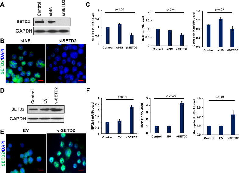
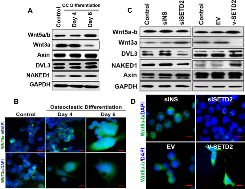
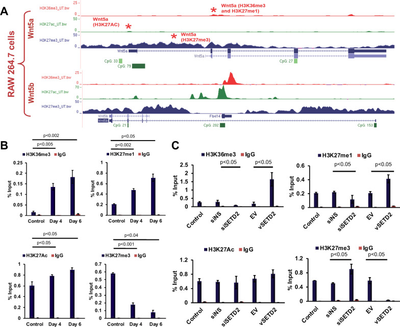
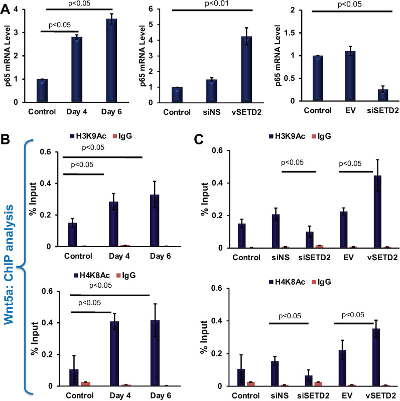
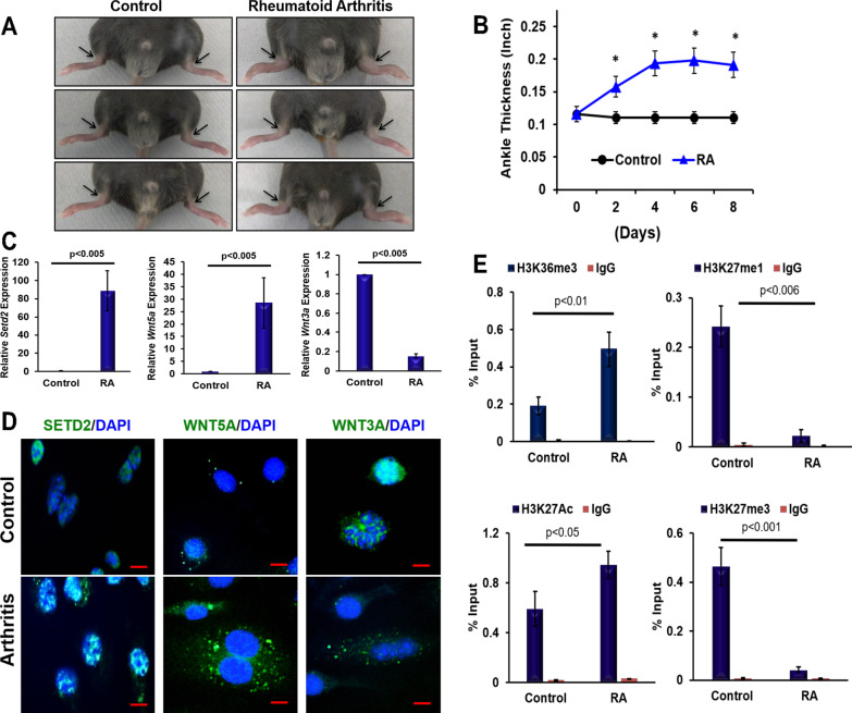
To define the role of SETD2 in the WNT5a signaling in the context of osteoclastogenesis, we exploited two different models: in vitro osteoclast differentiation, and K/BxN serum-induced arthritis model. We found that SETD2 and WNT5a were upregulated during osteoclast differentiation and after induction of arthritis. Using gain- and loss-of-function approaches in the myeloid cell, we confirmed that SETD2 regulated the osteoclast markers, and WNT5a via modulating active histone marks by enriching H3K36me3, and by reducing repressive H3K27me3 mark. Additionally, during osteoclastic differentiation, the transcription of Wnt5a was also associated with the active histone H3K9 and H4K8 acetylations. Mechanistically, SETD2 directed induction of NF-κβ expression facilitated the recruitment of H3K9Ac and H4K8Ac around the TSS region of the Wnt5a gene, thereby, assisting osteoclast differentiation. Together these findings for the first time revealed that SETD2 mediated epigenetic regulation of Wnt5a plays a critical role in osteoclastogenesis and induced arthritis. Model for the Role of SETD2 dependent regulation of osteoclastic differentiation. A In monocyte cells SETD2-dependent H3K36 trimethylation help to create open chromatin region along with active enhancer mark, H3K27Ac. This chromatin state facilitated the loss of a suppressive H3K27me3 mark. B Additionally, SETD2 mediated induction of NF-κβ expression leads to the recruitment of histone acetyl transferases, P300/PCAF, to the Wnt5a gene and establish H3K9Ac and H4K8Ac marks. Along with other activation marks, these acetylation marks help in Wnt5a transcription which leads to osteoclastogenesis.

Keywords: Epigenetic regulation; Osteoclastic differentiation; SETD2; Wnt signaling.


Conflict of interest statement

The authors declare no competing interests.

Figures

Fig. 1
Fig. 1
Elevated SETD2 expressions during osteoclastic differentiation. a TRAP staining was performed to detect differentiated osteoclasts at days 4 and 6. b Relative gene expression of Nfatc1, Trap, Cathepsin K, and Mmp9 were determined in differentiated cells using qRT-PCR keeping β-Actin as an internal control. c The relative gene expression of Setd2 was determined in differentiated cells. d Protein levels of SETD2 were detected by western blot and the protein expression level was analyzed by ImageJ software. The protein expression level was normalized by using GAPDH expression level and graphically represented. e Imunofluorescence analysis of SETD2 expression in osteoclast differentiated samples
Fig. 2
Fig. 2
SETD2 induced osteoclast markers expression. Levels of SETD2 was detected by western blot and immunofluorescence after siRNA-mediated knockdown (a, b, e), and overexpression (c, d, f) of SETD2 in RAW264.7 cells. Relative mRNA level expression of Nfatc1, Trap, and Cathepsin K were analyzed by qRT-PCR in SETD2 knocked down and overexpressed samples (e, f, respectively)
Fig. 3
Fig. 3
Canonical and non-canonical Wnt pathway molecules during osteoclastic differentiation, and after SETD2 overexpression and knockdown. a Western blot detection of Wnt signaling molecules during osteoclastic differentiation of RAW264.7 cells. b Detection of WNT5a and WNT3a molecules in cells during osteoclastic differentiation by immunofluorescence method. C. Western blot detection of Wnt signaling molecules after knockdown and overexpression of SETD2 in RAW264.7 cells. d Detection of WNT5A molecules in cells after overexpression and knockdown of SETD2 by immunofluorescence method. (Scale bar is 10 μM)
Fig. 4
Fig. 4
Transcriptional regulations of Wnt5a via SETD2. a UCSC genome browser analysis of RAW 264.7 cell line. The image is showing the enrichment pattern of H3K36me3, H3K27Ac, and H3K27me3 marks on the Wnt5a/b gene in wild type untreated RAW 264.7 cells. Red star (*) marks are specific regions that were analyzed by ChIP-qRT-PCR on the Wnt3a gene. b Quantitative RT-PCR analysis of ChIP-DNA for H3K36me3, H3K27me1, H3K27me3, and H3K27Ac enrichment on the Wnt5a gene using specific primers designed from the intragenic and upstream region of TSS in osteoclast differentiated cells. c ChIP was performed for H3K36me3, H3K27me1, H3K27me3, and H3K27Ac marks on the Wnt5agene in SETD2 knocked down and overexpressed cells. ChIP with IgG rabbit served as a negative control for all ChIP analyses
Fig. 5
Fig. 5
SETD2 facilitated the expression of NF-κβ thus H3K9Ac and H4K8Ac enrichment. a RT-PCR analysis of NF-κβ expression in osteoclast differentiated and SETD2 knocked down and overexpressed samples. b ChIP-qRT-PCR analysis of H3K9Ac and H4K8Ac enrichment on the Wnt5a gene in osteoclast differentiated and C. SETD2 knocked down and overexpressed cells. ChIP with IgG from rabbit served as a negative control for all ChIP analyses
Fig. 6
Fig. 6
K/BxN serum-induced arthritis effects on SETD2 and Wnt signaling molecules. a Ankle morphology (arrowhead) after induced arthritic development. b Measured ankle thickness is shown graphically after arthritic development. Star (*) indicates p < 0.05. c Relative gene expressions of Setd2, Wnt5a, and Wnt3a were determined in bone marrow monocytes after induction of arthritis in mice using qRT-PCR keeping β-Actin as an internal control. d Immunofluorescence analysis of SETD2, WNT5A, and WNT3A protein expression in bone marrow monocytes after arthritic development. e ChIP-qRT-PCR analysis of H3K36me3, H3K27me1, H3K27me3, and H3K27Ac enrichment on the Wnt5a gene in bone marrow monocytes after arthritic development. ChIP with IgG rabbit served as a negative control

References

    1. Firestein GS, McInnes IB. Immunopathogenesis of rheumatoid arthritis. Immunity. 2017;46(2):183–196. doi: 10.1016/j.immuni.2017.02.006. - DOI - PMC - PubMed
    1. Takayanagi H, Kim S, Koga T, Nishina H, Isshiki M, Yoshida H, Saiura A, Isobe M, Yokochi T, Inoue J, Wagner EF, Mak TW, Kodama T, Taniguchi T. Induction and activation of the transcription factor NFATc1 (NFAT2) integrate RANKL signaling in terminal differentiation of osteoclasts. Dev Cell. 2002;3(6):889–901. doi: 10.1016/S1534-5807(02)00369-6. - DOI - PubMed
    1. Soysa NS, Alles N. NF-kappaB functions in osteoclasts. Biochem Biophys Res Commun. 2009;378(1):1–5. doi: 10.1016/j.bbrc.2008.10.146. - DOI - PubMed
    1. Barski A, Cuddapah S, Cui K, Roh TY, Schones DE, Wang Z, Wei G, Chepelev I, Zhao K. High-resolution profiling of histone methylations in the human genome. Cell. 2007;129(4):823–837. doi: 10.1016/j.cell.2007.05.009. - DOI - PubMed
    1. Wagner EJ, Carpenter PB. Understanding the language of Lys36 methylation at histone H3. Nat Rev Mol Cell Biol. 2012;13(2):115–126. doi: 10.1038/nrm3274. - DOI - PMC - PubMed

Publication types

MeSH terms

LinkOut - more resources