. 2021:111:1-29.
doi: 10.1016/bs.aivir.2021.07.001. Epub 2021 Aug 23.

Parainfluenza virus entry at the onset of infection

Affiliations

Parainfluenza virus entry at the onset of infection

Tara C Marcink et al. Adv Virus Res. 2021.

Abstract

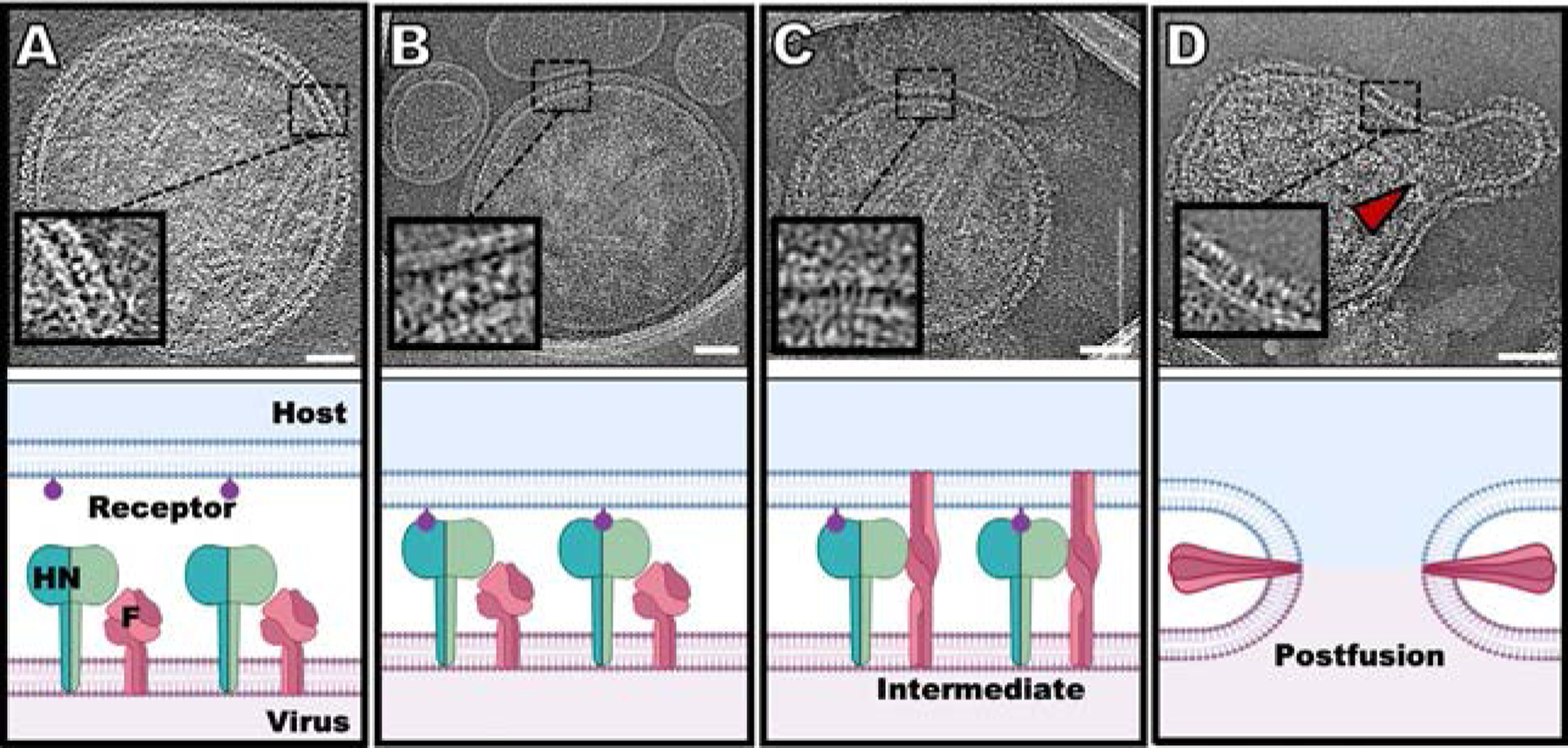
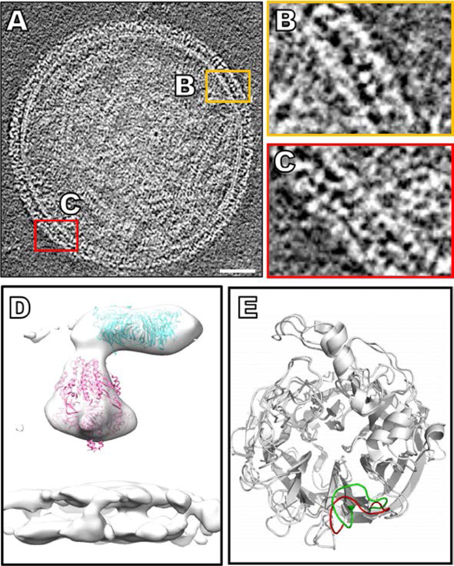
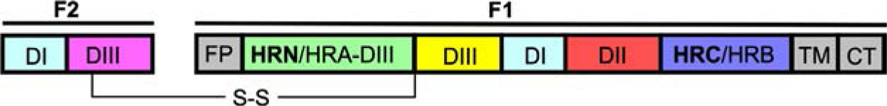
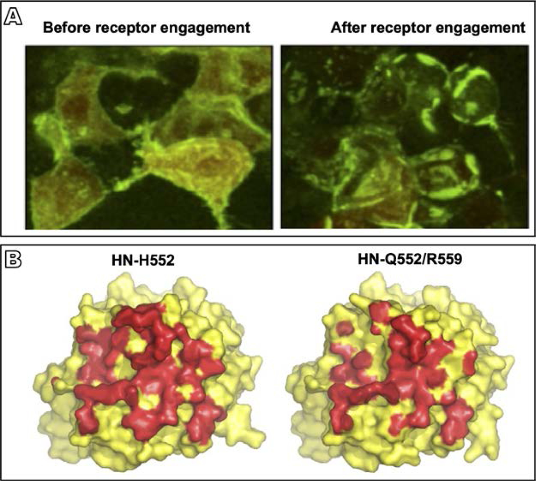
Parainfluenza viruses, members of the enveloped, negative-sense, single stranded RNA Paramyxoviridae family, impact global child health as the cause of significant lower respiratory tract infections. Parainfluenza viruses enter cells by fusing directly at the cell surface membrane. How this fusion occurs via the coordinated efforts of the two molecules that comprise the viral surface fusion complex, and how these efforts may be blocked, are the subjects of this chapter. The receptor binding protein of parainfluenza forms a complex with the fusion protein of the virus, remaining stably associated until a receptor is reached. At that point, the receptor binding protein actively triggers the fusion protein to undergo a series of transitions that ultimately lead to membrane fusion and viral entry. In recent years it has become possible to examine this remarkable process on the surface of viral particles and to begin to understand the steps in the transition of this molecular machine, using a structural biology approach. Understanding the steps in entry leads to several possible strategies to prevent fusion and inhibit infection.

Keywords: Antivirals; Cryo-electron tomography; Fusion inhibitors; Membrane fusion; Parainfluenza virus; Viral entry.


Figures

Fig. 1
Fig. 1
Schematic diagram of the steps in HPIV3 entry, with snapshots of cryo-electron tomographic images at each step (24). (A) HN (green) and F (dark pink) can be found densely packed on the viral surface (light pink). (B) Sialic acid (purple) binding to HN occurs in the presence of a surrogate host target membrane (erythrocyte membrane fragment) (blue). (C) Upon triggering of F by HN, F undergoes a large conformational change from a pre-fusion globular structure to a transient extended structure that crosses both membranes. (D) F folds back onto itself, pulling the viral and cell membranes toward each other, in a process that ultimately results in a merged membrane. Scale bars: (A–D) 50nm. Adapted from Marcink, T.C., Wang, T., des Georges, A., Porotto, M., Moscona A., 2020. Human parainfluenza virus fusion complex glycoproteins imaged in action on authentic viral surfaces. PLoS Pathog. 16 (9), e1008883, with permission.
Fig. 2
Fig. 2
The HN visualized on the virion surface and examined by cryo-electron tomography. (A) Contrast inverted cryo-ET central slice of HPIV3 before receptor engagement (Marcink et al., 2020b). (B and C) Enlarged regions of the surface glycoproteins with HN and F in tight apposition. (D) Sub-volume average of surface glycoproteins with crystal structure of the HN dimer (PDB ID: 4MZA) and the cryo-EM structure of pre-fusion F (PDB ID: 6MJZ) in green and pink (respectively), fitted into the sub-volume average of the HPIV3 viral surface. (E) A flexible loop on HN that adopts an open conformation (red) in direct vicinity of the active site of the apo-form of the protein and closes (green) upon inhibitor binding (Winger and von Itzstein, 2012). Panels (A–D): Adapted from Marcink, T.C., Wang, T., des Georges, A., Porotto, M., Moscona A., 2020. Human parainfluenza virus fusion complex glycoproteins imaged in action on authentic viral surfaces. PLoS Pathog. 16 (9), e1008883, with permission. Panel (E): From Winger, M., von Itzstein, M., 2012. Exposing the flexibility of human parainfluenza virus hemagglutinin-neuraminidase. J. Am. Chem. Soc. 134 (44), 18447–52, with permission.
Fig. 3
Fig. 3
Linear map of the HPIV fusion protein with fusion peptide (FP), transmembrane domain (TM), N-terminal heptad repeat (HRN/HRA-DIII) and C-terminal heptad repeat (HRC/HRB) domains.
Fig. 4
Fig. 4
HPIV3 HN-F interaction and the role of the HN dimer interface in this process. (A) 293T cells co-transfected with constructs encoding HN and F, each with their corresponding bimolecular fluorescence complementation tag, were treated overnight with zanamivir to prevent HN-receptor binding (Porotto et al., 2012a). Evenly distributed fluorescence was visualized across the cell surface in the absence of receptor engagement (left panel), but after the zanamivir was removed and receptor engagement was permitted, HN-F interaction occurred in clusters at the sites of cell-cell contact (right panel) (B) Surface rendering of the crystal structure of the HN monomer globular head viewed from above for HN with the indicated mutated site II residues (Xu et al., 2013). HN residues in contact with the opposing monomer in the dimer interface are shown in red. Introduction of the double mutation Q552/R559 in site II results in the loss of about 800 of 3700Å2 buried interface area in the dimer (right hand side), revealing the effect of alterations in this site to the dimer structure. Adapted from Porotto, M., Palmer, S.G., Palermo, L.M., Moscona, A., 2012a. Mechanism of fusion triggering by human parainfluenza virus type III: communication between viral glycoproteins during entry. J. Biol. Chem. 287 (1), 778–93, with permission; Xu, R., Palmer, S.G., Porotto, M., Palermo, L.M., Niewiesk, S., Wilson, I.A., et al., 2013. Interaction between the hemagglutinin-neuraminidase and fusion glycoproteins of human parainfluenza virus type III regulates viral growth in vivo, MBio. 4 (5), e00803–13, with permission.
Fig. 5
Fig. 5
The PIV F in its transient intermediate state during execution of fusion. (A) Examination of the pre-hairpin intermediate of PIV5 F by thin-sectioning and negative stain EM (Kim et al., 2011). Yellow arrows show intermediate F while the blue arrows show pre-fusion F distant from a region of interaction. (B) HPIV3 Contrast-inverted images where viral particles can be observed attached to target erythrocyte fragment membranes using cryo-ET (Marcink et al., 2020b). (C) Upper panel shows an enlarged region of interactions between the viral and target erythrocyte fragment membranes where elongated densities linking both membranes are visible. Insets include cyan lines where distance plot measurements were taken. Lower panel shows density line plots revealing a repeating 20–35Å-wide density. Scale bars: (B) 50nm. Panel (A): Adapted from Kim, Y.H., Donald, J.E., Grigoryan, G., Leser, G.P., Fadeev, A.Y., Lamb, R.A., et al., 2011. Capture and imaging of a prehairpin fusion intermediate of the paramyxovirus PIV5. Proc. Natl. Acad. Sci. U. S. A. 108 (52), 20992–7, with permission. Panels (B and C): From Marcink, T.C., Wang, T., des Georges, A., Porotto, M., Moscona A., 2020. Human parainfluenza virus fusion complex glycoproteins imaged in action on authentic viral surfaces. PLoS Pathog. 16 (9), e1008883, with permission.
Fig. 6
Fig. 6
Structural analysis of the mechanism of action of a small molecule that stimulates HN to trigger F. (A) Cells cotransfected with HN and F or transfected with F only were incubated in the presence of zanamivir alone to block HN-receptor binding, or with zanamivir together with CM9 or CSC11, two molecules that interact with HN to induce it to trigger F (Bottom-Tanzer et al., 2019). The cells were stained with monoclonal antibodies specific for the post-activated state of F. The proportions of activated F are shown as percentages of cells expressing post-triggered F on the y axis (±SD). (B) Overview of HPIV3 without and with PAC-3066, a next-generation small molecule that stimulates HN to activate F. Bars, 50nm (Marcink et al., 2020a). Lower panels show the viral surface glycoproteins without and with PAC-3066. (C) Cryo-electron 2D-subtomogram averages from a subset of viral surface glycoproteins in the absence of PAC-3066, where pre-fusion F can be identified (left) and with PAC3066, showing post-fusion F (Marcink et al., 2020a). Structural comparison coordinates of HN (PDB accession number 4MZA) and pre-fusion F (PDB accession number 6MJZ) are shown in pink and cyan, respectively. (D) Subtomogram averages where post-fusion F can be identified after PAC-3066 incubation. Structural comparison coordinates of the post-fusion F (PDB accession number 1ZTM) are shown in light blue. Panel (A): Adapted from Bottom-Tanzer, S.F., Rybkina, K., Bell, J.N., Alabi, C.A., Mathieu, C., Lu. M., et al., 2019. Inhibiting human parainfluenza virus infection by preactivating the cell entry mechanism. MBio 10 (1), with permission. Panels (B and C): From Marcink, T.C., Yariv, E., Rybkina, K., Mas, V., Bovier, F.T., des Georges, A., et al., 2020. Hijacking the fusion complex of human parainfluenza virus as an antiviral strategy, mBio 11 (1), with permission.
Fig. 7
Fig. 7
Neutralizing antibodies targeting pre-fusion F. (A) Structure-based design of prefusion-stabilizing mutations in PIV3 F with PIA174 antibody positioned at the apex of F (Stewart-Jones et al., 2018). HPIV3 F trimer domain organization showing DI (yellow), DII (red), HRN/HRA-DIII (green), F1-DIII (magenta), and HRC/HRB (blue). See Fig. 3 for description of domains. (B) Models of stabilized PIV1, PIV2, and PIV4, respectively, based on PIV5 pre-fusion F coordinates (PDB ID 4WSG) (Stewart-Jones et al., 2018). Boxes indicate regions of stabilizing mutations that were incorporated into F to generate pre-fusion F specific antibodies. Adapted from Stewart-Jones, G.B.E., Chuang, G.Y., Xu, K., Zhou, T., Acharya, P., Tsybovsky, Y., et al., 2018. Structure-based design of a quadrivalent fusion glycoprotein vaccine for human parainfluenza virus types 1–4. Proc. Natl. Acad. Sci. U. S. A. 115 (48), 12265–70, with permission.
Fig. 8
Fig. 8
Blocking fusion at the intermediate state of F. (A) HPIV3 F ectodomain in post-fusion conformation (PDB 1ZTM), with the inhibitory peptide shown coassembled with HPIV3-HRN underneath (PDB 6NRO) (Outlaw et al., 2020a). This inhibitory peptide mimics the post-fusion state blocking F in the intermediate state. (B left panel) Schematic of lipid-conjugated inhibitory peptide inserting into the target cell membrane via their lipid tails and “locking” the extended F in its transient intermediate state, preventing refolding to the post-fusion conformation (Marcink et al., 2020b). (B right panel) Contrast-inverted images where viral particles can be observed attached to target erythrocyte fragment membranes. Enlarged region of interaction between the viral and target erythrocyte fragment membranes show elongated densities linking both membranes. Panel (A): Adapted from Outlaw, V.K., Kreitler, D.F., Stelitano, D., Porotto, M., Moscona, A., Gellman, S.H., 2020. Effects of single alpha-to-beta residue replacements on recognition of an extended segment in a viral fusion protein. ACS Infect. Dis., with permission. Panel (B): From Marcink, T.C., Wang, T., des Georges, A., Porotto, M., Moscona A., 2020. Human parainfluenza virus fusion complex glycoproteins imaged in action on authentic viral surfaces. PLoS Pathog. 16 (9), e1008883, with permission.

References

    1. Bagai S, Lamb R, 1995. Quantitative measurement of paramyxovirus fusion: differences in requirements of glycoproteins between SV5 and human parainfluenza virus 3 or Newcastle disease virus. J. Virol 69, 6712–6719. - PMC - PubMed
    1. Bailly B, Dirr L, El-Deeb IM, Altmeyer R, Guillon P, von Itzstein M, 2016. A dual drug regimen synergistically blocks human parainfluenza virus infection. Sci. Rep 6, 24138. - PMC - PubMed
    1. Benhaim MA, Mangala Prasad V, Garcia NK, Guttman M, Lee KK, 2020. Structural monitoring of a transient intermediate in the hemagglutinin fusion machinery on influenza virions. Sci. Adv 6 (18), eaaz8822. - PMC - PubMed
    1. Benton DJ, Gamblin SJ, Rosenthal PB, Skehel JJ, 2020. Structural transitions in influenza haemagglutinin at membrane fusion pH. Nature 583 (7814), 150–153. - PMC - PubMed
    1. Bishop KA, Hickey AC, Khetawat D, Patch JR, Bossart KN, Zhu Z, et al., 2008. Residues in the stalk domain of the hendra virus g glycoprotein modulate conformational changes associated with receptor binding. J. Virol 82 (22), 11398–11409. - PMC - PubMed

Substances

LinkOut - more resources