Review
. 2021 Oct;26(4):379-384.
doi: 10.17712/nsj.2021.4.20210081.

Invasive aspergillosis of the central nervous system in immunocompetent patients in Saudi Arabia: Case series and review of the literature

Affiliations
Review

Invasive aspergillosis of the central nervous system in immunocompetent patients in Saudi Arabia: Case series and review of the literature

Mahmoud S Taha et al. Neurosciences (Riyadh). 2021 Oct.

Abstract

Objective: Invasive aspergillosis of the central nervous system in immunocompetent patients is a rare disease. We present in this study three cases that were treated in our centre and reviewed the results of similar studies from Saudi Arabia.

Methods: We retrospectively reviewed all cases of invasive aspergillosis of the central nervous system (CNS) that were treated in our hospital in the last 10 years. We also reviewed the literature for any similar series published from Saudi Arabia.

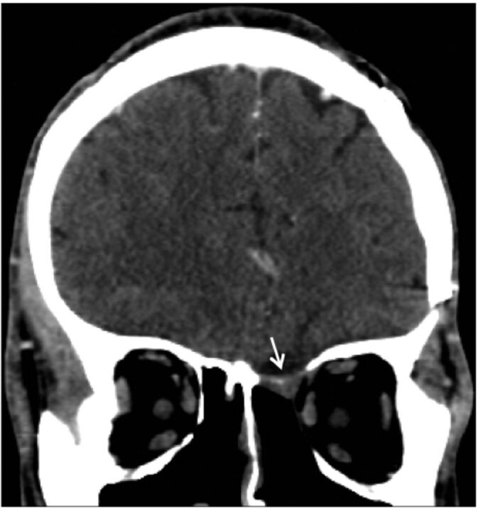
Results: We had three cases treated in our centre and we also found three similar case series in the literature. Total number of cases, including our series was 28, age range from 17 to 66, 10 men and 18 women. The source of infection was nasal sinuses in all cases. Initial presentations were variable and included headache (80% of cases), proptosis or diplopia (50% of cases), seizures (20% of cases), cranial nerve palsies (18% of cases) and acute deterioration in level of consciousness (18% of cases). All patients underwent surgery followed by long course of antifungal treatment. Clinical outcome was reported as cured or no recurrence in 13 cases (47%).

Conclusions: Invasive aspergillosis of CNS is a rare disease in immunocompetent patients. Despite treatment prognosis remains unfavourable in many cases.


Figures

Figure 1
Figure 1
- Axial enhanced brain computed tomography (CT) A) Axial enhanced brain computed tomography (CT) shows hyperdense opacification of the right frontal paranasal sinus with erosion of its posterior wall (black dashed arrow), B) Brain magnetic resonance imaging MRI scan Axial T2-weighted and, C) MRI T1-weighted post contrast show a large intra-axial lesion in the right frontal lobe with irregular thick peripheral enhancement (white arrows), D) MRI diffusion-weighted imaging shows no corresponding central diffusion restriction.
Figure 2
Figure 2
- Hematoxylin- and eosin-stained sections (magnification, 40x) showing A) neuroglial tissue containing ill-defined granulomata with multinucleated giant cells. Thin hyphae with regular septation and branching at a 45° angle are noted, B) Grocott’s methenamine silver stain demonstrating thin hyphae and spores (magnification, 20x).
Figure 3
Figure 3
- Detection methods for mycology: Specimens were inoculated on both selective fungal media (Mycosel, Becton, Dickenson and Company, MD, USA) and general fungal media Sabouraud Dextrose Agar (Titan Biotech Ltd, India) and incubated at 28°C–30°C for up to 4 weeks. Cultures with fungal growth were further worked up for mould identification using tease mount, scotch tape preparation and potato dextrose agar media for conidiation.The culture grew rapidly, producing a green–grey colony. The mould produced septate hyaline hyphae with smooth conidiophores supporting a dome-shaped vesicle. A row of phialides on the upper surface of the vesicle bore chains of round conidia in a columnar fashion characteristic of Aspergillus fumigatus.
Figure 4
Figure 4
- Axial plain brain CT shows (A) post-craniotomy changes in the right frontal aspect of the skull with haemorrhagic collection within the surgical bed (white arrow). Extensive ischemic low-attenuation tissue swelling in the right cerebral hemisphere and left frontal lobe is also observed (asterisks) along with a left-sided midline shift. (B) 3-dimensional reformatted MIP image from CTA shows occlusion of the right M1 segment (dashed arrow) and bilateral ACA (bidirectional arrow).
Figure 5
Figure 5
- Enhanced coronal brain CT shows a growing mass centred in the anterior skull base with bone erosions (arrows) along with intracranial, extra-axial and ethmoid sinus soft tissue components.
Figure 6
Figure 6
- Enhanced coronal brain CT on post-operative follow up does not show any evidence of recurrence.

References

    1. Patterson TF, Thompson, GR, Denning DW, Fishman JA, Hadley S, Herbrecht R, et al. . 2016. Practice guidelines for the diagnosis and management of aspergillosis: 2016 Update by the Infectious Diseases Society of America. Clin Infect Dis 2016; 63: e1–e60. - PMC - PubMed
    1. Miceli, MH. Central nervous system infections due to aspergillus and other hyaline molds. J Fungi (Basel) 2019; 5: 79. - PMC - PubMed
    1. Siddiqui AA, Shah AA, Bashir SH.. Craniocerebral aspergillosis of sinonasal origin in immunocompetent patients: clinical spectrum and outcome in 25 cases. Neurosurgery 2004; 55: 602–11; discussion 611-613. - PubMed
    1. Nadkarni T, Goel A.. Aspergilloma of the brain: an overview. J Postgrad Med 2005; 51: S37–S41. - PubMed
    1. Al-Bhlal LA. Fungal infection of the nasal cavity and paranasal sinuses: Review of 26 cases. Ann Saudi Med 1996; 16: 615–621. - PubMed

Substances

LinkOut - more resources