Review
. 2022 Jan;35(Suppl 1):36-50.
doi: 10.1038/s41379-021-00943-2. Epub 2021 Oct 18.

Lung neuroendocrine neoplasms: recent progress and persistent challenges

Affiliations
Review

Lung neuroendocrine neoplasms: recent progress and persistent challenges

Natasha Rekhtman. Mod Pathol. 2022 Jan.

Abstract

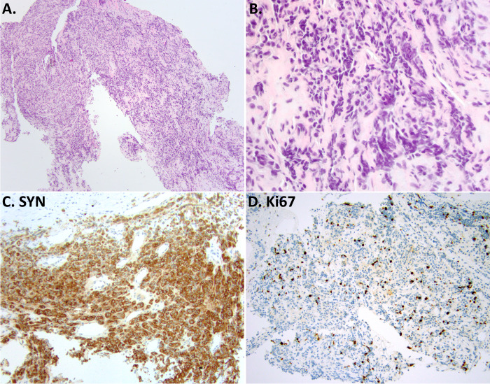
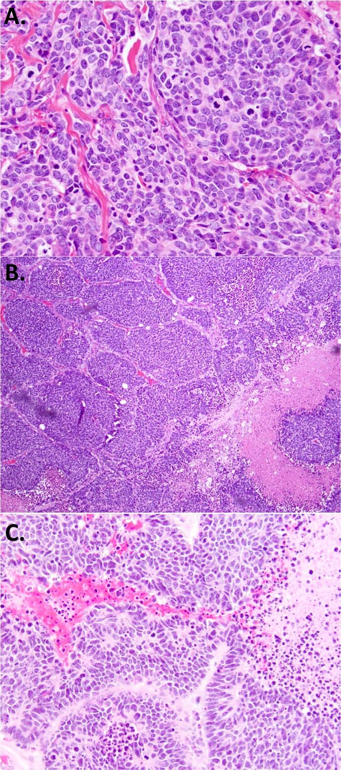
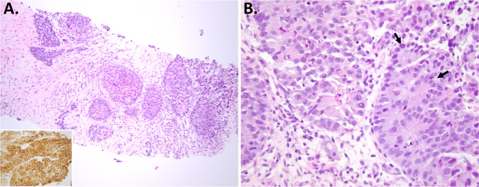
This review summarizes key recent developments relevant to the pathologic diagnosis of lung neuroendocrine neoplasms, including carcinoids, small cell lung carcinoma (SCLC), and large cell neuroendocrine carcinoma (LCNEC). Covered are recent insights into the biological subtypes within each main tumor type, progress in pathological diagnosis and immunohistochemical markers, and persistent challenging areas. Highlighted topics include highly proliferative carcinoids and their distinction from small cell and large cell neuroendocrine carcinomas (NECs), the evolving role of Ki67, the update on the differential diagnosis of NEC to include thoracic SMARCA4-deficient undifferentiated tumors, the recent data on SCLC transcriptional subtypes with the emergence of POU2F3 as a novel marker for the diagnosis of SCLC with low/negative expression of standard neuroendocrine markers, and the update on the diagnosis of LCNEC, particularly in biopsies. There has been remarkable recent progress in the understanding of the genetic and expression-based profiles within each type of lung neuroendocrine neoplasm, and it is hoped that these insights will enable the development of novel diagnostic, prognostic, and predictive biomarkers to aid in the pathologic assessment of these tumors in the future.


Conflict of interest statement

The author declares no competing interests.

Figures

Fig. 1
Fig. 1. Example of a highly proliferative carcinoid (current WHO terminology “LCNEC with morphologic features of carcinoid tumor”).
Example from 2 tumors (A, B tumor 1, CF tumor 2). H&E images show cytologically land tumors, with uniform nuclei and smooth nuclear membranes, typical of carcinoids/NETs, but with strikingly elevated mitotic counts indicated by arrows (>10 per 2 mm2). Synaptophysin (SYN; D) is strongly and diffusely positive. Ki67 is heterogenous at low-power (E) but in hot-spot areas exceeds 30% (F). Case 1 corresponds to case ID LCNEC-36 in Rekhtman et al., which was found to harbor an MEN1 mutation.
Fig. 2
Fig. 2. Ki67 for the diagnosis of carcinoid in a crushed biopsy.
Low-power (A) and high-power (B) images of bronchoscopic biopsy, where crush artifact leads to the appearance of cell molding, and together with positive synaptophysin (SYN; C) closely mimics the appearance of SCLC. Mitoses are not evaluable in such specimens, but the low Ki67 index (<5%) readily identifies this as a crushed carcinoid rather than SCLC. This remains a common diagnostic pitfall.
Fig. 3
Fig. 3. Morphologic features in SCLC that may be misinterpreted as LCNEC.
A SCLC with larger overall cell size and more abundant cytoplasm, but otherwise morphologic features fitting the diagnosis of SCLC (notice there are no visible intercellular borders). B SCLC commonly have at least focal nested architecture; nested pattern alone does not favor the diagnosis of LCNEC over SCLC. C Rosettes are also common in SCLC.
Fig. 4
Fig. 4. POU2F3 expression in SCLC with minimal reactivity for standard NE markers.
Low-power (A) and high-power (B) images showing morphologic appearance suggestive of SCLC, but with completely negative labeling for synaptophysin (SYN; C), chromogranin A (CHGA, D), and INSM1 (F). Only CD56—the least specific NE marker—shows labeling in scattered cells (E). The diagnosis of SCLC is supported by the strong expression of POU2F3. As expected for small cell carcinoma, Rb shows the loss of expression in tumor cells relative to entrapped benign cells (G). Photo credit: Marina K Baine.
Fig. 5
Fig. 5. Thoracic SMARCA4-deficient undifferentiated tumor as a mimicker of SCLC.
Low-power (A) and higher-power (B) histologic images of highly necrotic round cell tumor with crush artifact and molding, which in conjunction with synaptophysin labeling (SYN, C) closely mimics SCLC. In better-preserved areas, prominent nucleoli and rhabdoid cells were seen (not shown). The diagnosis is confirmed by the loss of SMARCA4 (BRG1) expression in tumor cells relative to entrapped benign cells (D). Other markers supporting the diagnosis include SMARCA2 (BRM) co-deficiency and the expression of stem-cell markers (SALL4, CD34).
Fig. 6
Fig. 6. Prototypical morphologic spectrum in LCNEC.
Panel A illustrates LCNEC morphology falling in the differential diagnosis with NSCLC. Basaloid appearance, with rosettes and palisading, together with robust expression of NE markers supports the diagnosis. Panel B illustrates two LCNECs with morphology falling in the differential diagnosis with SCLC—cells are more crowded with molding but show obvious nucleoli and visible intercellular membranes.
Fig. 7
Fig. 7. Example of LCNEC diagnosis in a core biopsy.
Low-power (A) and high-power (B) images show a clear-cut NSCLC but with NE architecture (trabeculae, palisaded nests, rosettes), with high proliferation rate, extensive necrosis, and diffuse expression of NE markers (SYN shown in inset in A). Biopsy of this type can be diagnosed as definite LCNEC. Arrows in B mark rosettes.

References

    1. WHO Classification of Tumours Editorial Board. Thoracic Tumours. 5th ed. Lyon (France): International Agency for Research on Cancer. (2021).
    1. Rekhtman N. Neuroendocrine tumors of the lung: an update. Arch. Pathol. Lab. Med. 2010;134:1628–1638. - PubMed
    1. Seidel D, et al. A genomics-based classification of human lung tumors. Sci. Transl. Med. 2013;5:1–15. - PMC - PubMed
    1. Cree, I. A. et al. Counting mitoses: SI(ze) matters! Modern Pathol.10.1038/s41379-021-00825-7 (2021). - PMC - PubMed
    1. WHO Classification of Tumours Editorial Board. Digestive System Tumours. 5th ed. Lyon (France): International Agency for Research on Cancer. (2019).

Publication types

MeSH terms

Substances