. 2021 Dec;125(12):1666-1676.
doi: 10.1038/s41416-021-01560-1. Epub 2021 Oct 18.

Retrospective analysis of Schlafen11 (SLFN11) to predict the outcomes to therapies affecting the DNA damage response

Affiliations

Retrospective analysis of Schlafen11 (SLFN11) to predict the outcomes to therapies affecting the DNA damage response

Sophie E Willis et al. Br J Cancer. 2021 Dec.

Abstract

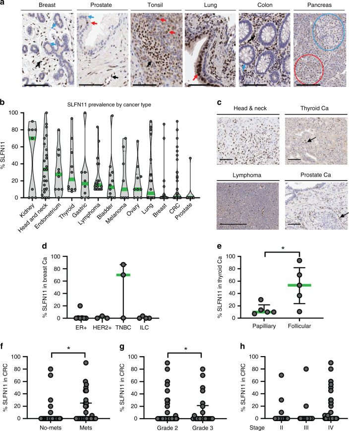
Background: The absence of the putative DNA/RNA helicase Schlafen11 (SLFN11) is thought to cause resistance to DNA-damaging agents (DDAs) and PARP inhibitors.

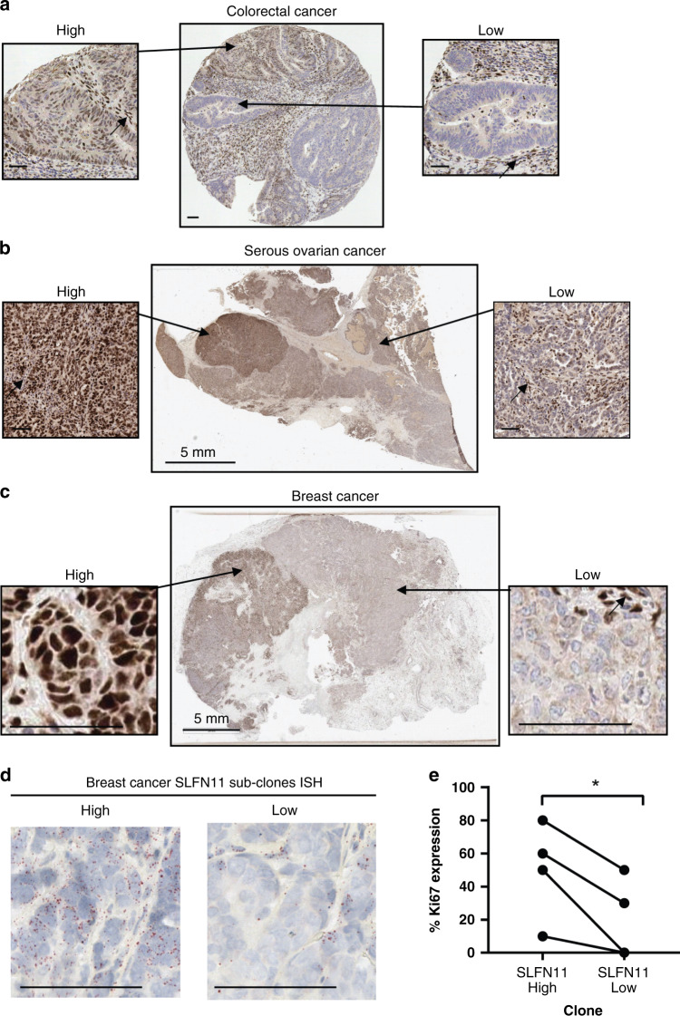
Methods: We developed and validated a clinically applicable SLFN11 immunohistochemistry assay and retrospectively correlated SLFN11 tumour levels to patient outcome to the standard of care therapies and olaparib maintenance.

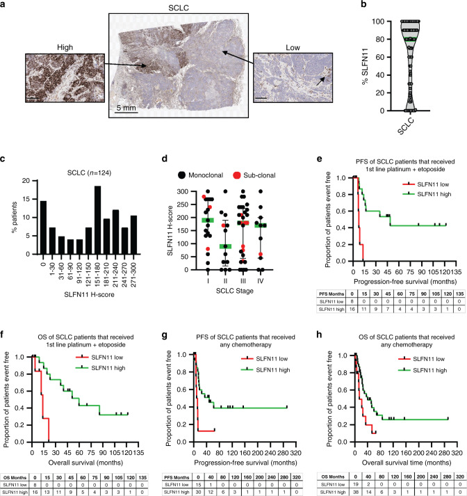
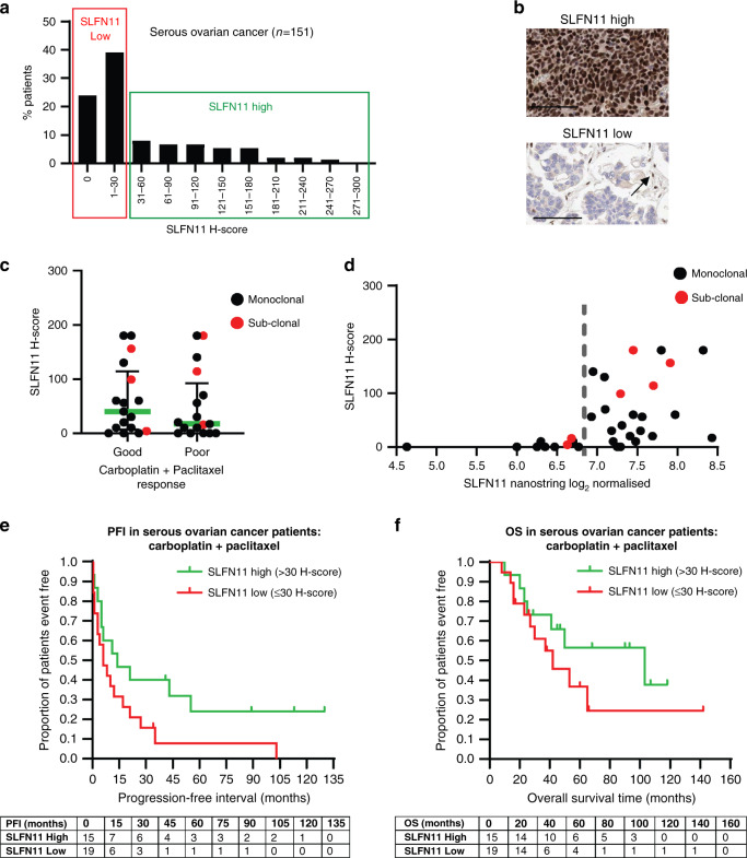
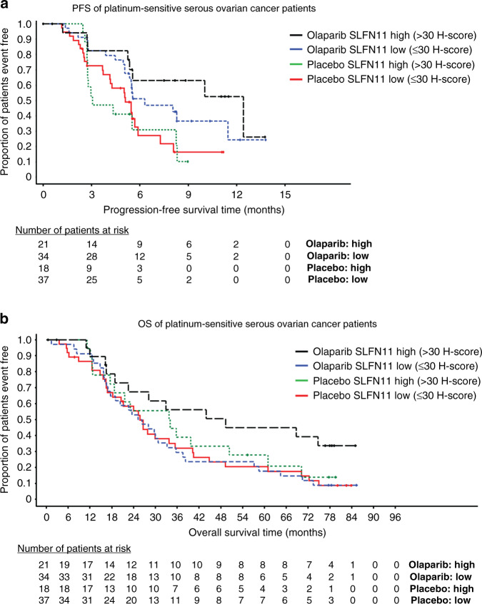
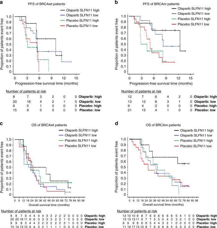
Results: High SLFN11 associated with improved prognosis to the first-line treatment with DDAs platinum-plus-etoposide in SCLC patients, but was not strongly linked to paclitaxel-platinum response in ovarian cancer patients. Multivariate analysis of patients with relapsed platinum-sensitive ovarian cancer from the randomised, placebo-controlled Phase II olaparib maintenance Study19 showed SLFN11 tumour levels associated with sensitivity to olaparib. Study19 patients with high SLFN11 had a lower progression-free survival (PFS) hazard ratio compared to patients with low SLFN11, although both groups had the benefit of olaparib over placebo. Whilst caveated by small sample size, this trend was maintained for PFS, but not overall survival, when adjusting for BRCA status across the olaparib and placebo treatment groups, a key driver of PARP inhibitor sensitivity.

Conclusion: We provide clinical evidence supporting the role of SLFN11 as a DDA therapy selection biomarker in SCLC and highlight the need for further clinical investigation into SLFN11 as a PARP inhibitor predictive biomarker.


Conflict of interest statement

SEW, CW, MPR, TB, PMC, EVJ, PR, JRC, HKA, FSLN, PMW, DH, ED, EAH, JCB, AJP, EL & GNJ were employees of AstraZeneca during this study. All other authors declare no competing interests.

Figures

Fig. 1
Fig. 1. SLFN11 protein levels are variable across tumour types and higher levels are found in more aggressive cancer subtypes.
a SLFN11 expression in normal tissues, arrows indicate (from left to right); negative breast lobular cells, positive (red) and negative (blue) prostatic cells, positive lymphocytes, positive bronchial epithelial cells, negative colon epithelial cells. Negative Langerhans islets (blue circle) and positive cluster of acinar cells (red circle) also shown. b SLFN11 prevalence in various cancer types from multi-tumour tissue microarrays (TMAs), expression of SLFN11 shown as percent of positive tumour cells when internal control staining was acceptable. Median given (green) for each cancer type. c Example images of SLFN11 IHC staining in head and neck, thyroid, lymphoma and prostate cancers (Ca). d, e Breakdown of the SLFN11 protein levels by: d breast cancer subtype; oestrogen receptor-positive (ER + ), human epidermal growth factor receptor 2 (HER2 + ), triple-negative breast cancer (TNBC), invasive lobular breast cancer (ILC) and e thyroid cancer subtype; papillary and follicular. fh SLFN11 expression in colorectal cancer (CRC) patients (n = 144) subcategorised by clinical patient information: f presence of metastasis or not; g tumour grade, h disease stage. Data shown as median ± interquartile range. *P value <0.05 by Mann–Whitney test. Black arrows are stromal and endothelial cells. Scale bars at 100 µm.
Fig. 2
Fig. 2. SLFN11 protein expression in clinical tissues can be sub-clonal, with both high- and low-expressing regions within a single tumour.
ac SLFN11 sub-clonal expression pattern demonstrated by IHC of a a CRC TMA core, b resected serous ovarian cancer tissue, c resected breast cancer tissue. A magnified image of the high- and low SLFN11 subclones are shown for each. Black arrows indicate stromal and endothelial cells used as a positive internal control. d RNA in situ hybridisation (ISH) of SLFN11 showing expression of SLFN11 RNA transcripts in the breast cancer SLFN11 subclones. e SLFN11 sub-clonal expression correlated to Ki67 expression. Line indicates patient-matched subclones. *P value < 0.05 paired t test. Scale bars at 50 µm unless otherwise stated.
Fig. 3
Fig. 3. High SLFN11 in small-cell lung cancer (SCLC) patients treated with standard of care linked to improved clinical outcome.
a Image of a SCLC patient tumour demonstrating sub-clonal SLFN11 expression, areas from the high- and low-expressing subclones are magnified. The black arrow indicates SLFN11-positive stromal cells in the negative sub-clone. Scale bars at 100 µm unless otherwise stated. b SLFN11 prevalence by IHC in SCLC patients, median in green. c Bimodal prevalence of SLFN11 in the SCLC cohort (n = 124). d SLFN11 expression in SCLC subcategorised by disease stage. Black; SLFN11 monoclonal expression, Red; SLFN11 sub-clonal tumours. Median ± interquartile range shown. eh Kaplan–Meier analysis of SCLC patients categorised by SLFN11 expression; SLFN11 high (H-score >122) compared to low (H-score ≤122). e Progression-free survival (PFS) and f overall survival (OS) in months for patients (n = 24) that received first-line platinum (carboplatin or cisplatin) plus etoposide. g PFS and h OS in months for 45 and 57 patients, respectively, that received any chemotherapy. Events table with patients at risk shown for each timepoint.
Fig. 4
Fig. 4. SLFN11 not significantly associated with sensitivity to the paclitaxel–carboplatin doublet in serous ovarian cancers.
a Prevalence of SLFN11 by IHC H-score in serous ovarian cancer patient biopsies (n = 151), cut-off depicting the high and low SLFN11 subgroups shown, b representative images of high and low SLFN11 tumours. Black arrow indicates SLFN11-positive stromal cells used as an internal control. Scale bars at 100 µm. c SLFN11 H-score of patients divided by extreme good and poor clinical responders to carboplatin and paclitaxel doublet therapy. Median ± interquartile range shown. d SLFN11 IHC protein expression by H-score compared to NanoString quantified SLFN11 gene expression in the HGSOC patients. Dotted line indicates gene expression threshold that distinguishes positive from negative SLFN11 patients. Patients with sub-clonal SLFN11 indicated by a red dot. e, f Kaplan–Meier survival curves of (e) progression-free interval (PFI) and (f) overall survival (OS) of high (>30 H-score SLFN11) vs. low SLFN11 ( ≤ 30 H-score SLFN11) subgroups in the 34 patients with high-grade serous ovarian cancer treated with carboplatin and paclitaxel doublet. Events table with patients at risk shown for each timepoint.
Fig. 5
Fig. 5. Elevated SLFN11 levels in HGSOC patients confers sensitivity to olaparib.
a Cox proportional hazards model of progression-free survival (PFS) of high (>30 H-score SLFN11) vs. low SLFN11 ( ≤ 30 H-score SLFN11) subgroups in platinum-sensitive serous ovarian carcinoma patients treated with either placebo or olaparib. b Overall survival (OS) of high vs. low SLFN11 in the same study. Events table with patients at risk shown for each timepoint.
Fig. 6
Fig. 6. SLFN11 does not predict olaparib sensitivity independent of BRCA status.
ad Cox proportional hazards model by SLFN11 high (>30 H-score SLFN11) vs. low SLFN11 ( ≤ 30 H-score SLFN11) subgroups in platinum-sensitive serous ovarian carcinoma patients treated with either placebo or olaparib for (a) progression-free survival (PFS) in BRCA wild-type patients, b PFS in BRCA mutant patients, c Overall survival (OS) in BRCA wild-type patients, d OS in BRCA mutant patients. Events table with patients at risk shown for each timepoint. VUS BRCAm patients included in the BRCAwt group.

References

    1. Murai J, Thomas A, Miettinen M, Pommier Y. Schlafen 11 (SLFN11), a restriction factor for replicative stress induced by DNA-targeting anti-cancer therapies. Pharm Ther. 2019;201:94–102. doi: 10.1016/j.pharmthera.2019.05.009. - DOI - PMC - PubMed
    1. Hopkins TA, Ainsworth WB, Ellis PA, Donawho CK, DiGiammarino EL, Panchal SC, et al. PARP1 trapping by PARP inhibitors drives cytotoxicity in both cancer cells and healthy bone marrow. Mol Cancer Res. 2019;17:409–19.. doi: 10.1158/1541-7786.MCR-18-0138. - DOI - PubMed
    1. Iwasaki J, Komori T, Nakagawa F, Nagase H, Uchida J, Matsuo K, et al. Schlafen11 expression is associated with the antitumor activity of trabectedin in human sarcoma cell lines. Anticancer Res. 2019;39:3553–63.. doi: 10.21873/anticanres.13501. - DOI - PubMed
    1. Nogales V, Reinhold WC, Varma S, Martinez-Cardus A, Moutinho C, Moran S, et al. Epigenetic inactivation of the putative DNA/RNA helicase SLFN11 in human cancer confers resistance to platinum drugs. Oncotarget. 2016;7:3084–97. doi: 10.18632/oncotarget.6413. - DOI - PMC - PubMed
    1. Shee K, Wells JD, Jiang A, Miller TW. Integrated pan-cancer gene expression and drug sensitivity analysis reveals SLFN11 mRNA as a solid tumor biomarker predictive of sensitivity to DNA-damaging chemotherapy. PLoS ONE. 2019;14:e0224267. doi: 10.1371/journal.pone.0224267. - DOI - PMC - PubMed

Publication types