Review
. 2021 Dec;22(12):758-776.
doi: 10.1038/s41583-021-00526-9. Epub 2021 Oct 18.

A neuropeptide code for itch

Affiliations
Review

A neuropeptide code for itch

Zhou-Feng Chen. Nat Rev Neurosci. 2021 Dec.

Abstract

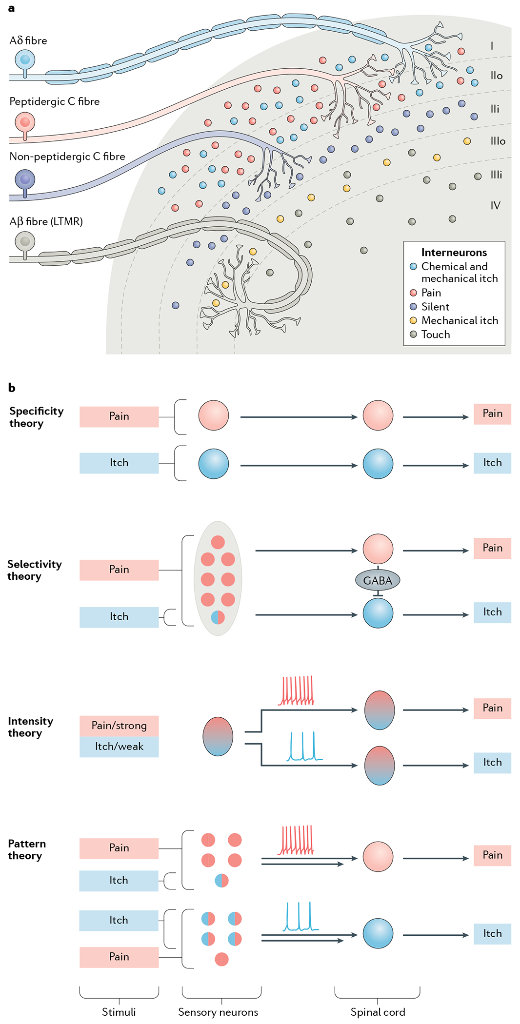
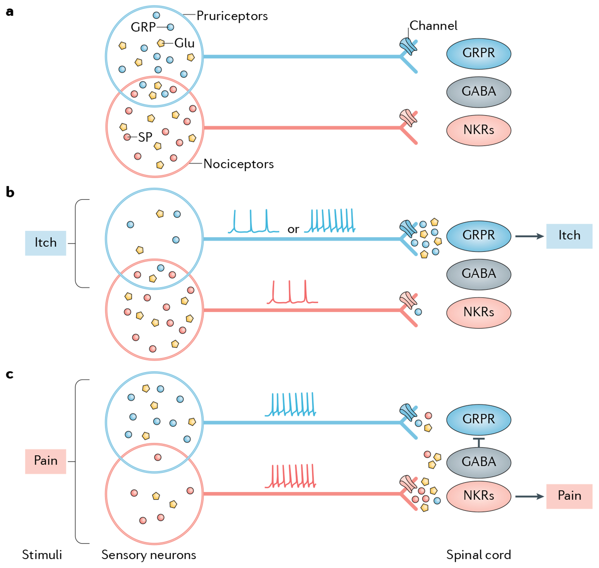
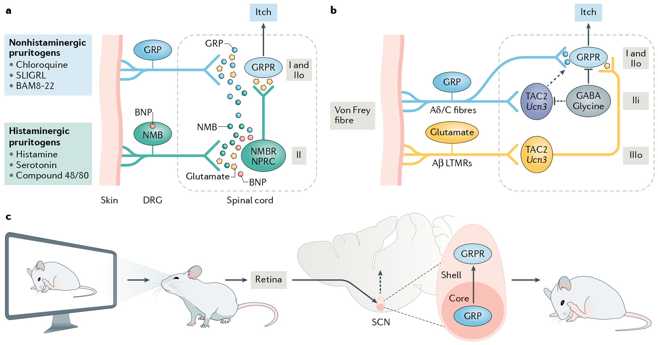
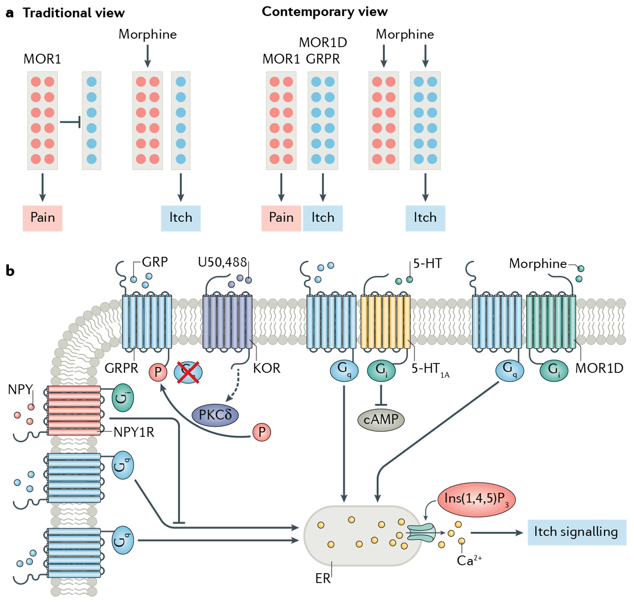
Itch is one of the most primal sensations, being both ubiquitous and important for the well-being of animals. For more than a century, a desire to understand how itch is encoded by the nervous system has prompted the advancement of many theories. Within the past 15 years, our understanding of the molecular and neural mechanisms of itch has undergone a major transformation, and this remarkable progress continues today without any sign of abating. Here I describe accumulating evidence that indicates that itch is distinguished from pain through the actions of itch-specific neuropeptides that relay itch information to the spinal cord. According to this model, classical neurotransmitters transmit, inhibit and modulate itch information in a context-, space- and time-dependent manner but do not encode itch specificity. Gastrin-releasing peptide (GRP) is proposed to be a key itch-specific neuropeptide, with spinal neurons expressing GRP receptor (GRPR) functioning as a key part of a convergent circuit for the conveyance of peripheral itch information to the brain.


Conflict of interest statement

Competing interests

The author declares no competing interests.

Figures

Fig. 1 |
Fig. 1 |. Dorsal horn architecture and coding theories for itch.
a | The dorsal horn, illustrating its division into several distinct laminae (I–IV). Laminae I and II are innervated by unmyelinated C fibres and fast-conducting myelinated Aδ fibres, which together convey information relevant to pain, itch, pleasant touch and temperature sensing. Interneurons in laminae I and II additionally receive mechanical itch information from Aβ low-threshold mechanoreceptors (LTMRs) via an indirect route (not shown),. Lamina II can be subdivided into two layers: unmyelinated peptidergic C fibres terminate mainly in lamina I and the outer layer of lamina II and convey information that requires slow conduction velocities, such as that important for chemical itch and chemical pain,. The inner layer of lamina II is innervated mainly by unmyelinated non-peptidergic C fibres that convey pain, itch and other somatosensory information. Laminae III and IV are typically innervated by myelinated Aβ LTMR fibres that convey mechanical touch, mechanical itch and proprioceptive information,,,,. Lamina III can be further divided into two layers. The outer layer contains TAC2 neurons, which mediate mechanical itch. Dorsal horn projection neurons in lamina I and possibly laminae III and IV, integrate and relay itch information to the somatosensory cortex (not shown),,. b | The main current coding theories of itch. According to the specificity (or labelled line) theory, information about itch-inducing and painful stimuli is conveyed via distinct neural pathways from the skin to the spinal cord. There are itch-specific neurons in the periphery, which are activated by itch stimuli exclusively.The selectivity (or population coding) theory proposes that pruriceptors (represented by a circle filled half in red and half in blue) are a small subset of nociceptive neurons. Pruriceptors respond to both itch-inducing and painful stimuli, whereas a much larger population of nociceptors (red) convey pain information exclusively3,19. This theory suggests that distinct labelled lines for pain and itch exist in the spinal cord and that pain transmission can activate spinal inhibitory neurons to inhibit or occlude itch by crosstalk between the two labelled lines,. According to the intensity theory, weak activation of nociceptors provokes itch, whereas strong activation of the same population of neurons evokes pain, via different modes of firing pattern (tonic for weak and burst for strong stimuli),,,. Like the selectivity theory, the pattern theory, postulates that within the larger population of sensory nociceptive neurons (red) there is a subset of pruriceptors (half red, half blue). However, it suggests that the distribution of these pruriceptors may be different in the epidermis and dermis (not shown). Therefore, depending on the nature and location of a given stimulus, a mixture of pruriceptors and nociceptors will be differentially activated, leading to different population firing patterns (tonic versus burst) and thereby encoding itch or pain specificity,. This theory does not explain how itch and pain information is encoded in the spinal cord. However, it can be assumed that itch and pain are conveyed separately in the spinal cord after the encoding by primary sensory neurons.
Fig. 2 |
Fig. 2 |. Hypothetical model for the neuropeptide coding of itch and pain.
The schematics illustrate the neuropeptide code model, which suggests that itch and pain are encoded and transmitted separately through discrete neuropeptides. Substance P (SP) is shown as an example of a pain-specific neuropeptide transmitter, whereas gastrin-releasing peptide (GRP) is shown as an example of an itch-specific neuropeptide transmitter. Other types of itch-specific or pain-specific neuropeptides may exist but are not shown for illustrative purposes (see also FiG. 3). a | Pruriceptors and nociceptors partially overlap, which is manifested in co-expression of GRP and SP. In the absence of itch or pain stimuli, neither GRP nor SP is released from its terminal, resulting in a lack of itch or pain transmission. For simplistic purposes, neurotransmitters are shown only in the cell bodies of nociceptors and pruriceptors. b | Under itchy conditions, pruriceptors are activated either spontaneously (resulting in tonic firing) or in response to itch stimuli (resulting in bursting firing). This activation is proposed to release GRP and glutamate into the spinal cord, activating GRP receptor (GRPR)-expressing neurons (GRPR neurons), which transmit itch information exclusively to the brain. A smaller number of nociceptors that co-express GRP and SP (indicated as the overlap of the ovals representing these two cell populations) are also activated but fail to release SP, owing to tight control of presynaptic neurotransmitter release via channels such as sodium or calcium channels. Therefore, no pain information is transmitted. This subset of neurons may also release a small amount of GRP at synapses with spinal nociceptive neurons expressing NK receptors (NKRs), but will fail to activate them owing to the lack of GRPR in these neurons. The GABAergic inhibitory interneurons that innervate itch-specific spinal neurons remain inactive. c | Under painful conditions, a population of nociceptors that contain some pruriceptors are concomitantly activated via a bursting firing pattern that provokes the release of SP and glutamate from these neurons,,, which activate spinal nociceptive neurons expressing NKRs. A small amount of GRP may be co-released but fails to activate these neurons owing to the lack of GRPR expression. Meanwhile, a smaller subset of pruriceptors expressing both GRP and SP or GRP alone (not shown) (indicated as the overlap of the ovals representing these two cell populations) is also activated and releases GRP and glutamate, which marginally activate GRPR neurons. A small amount of SP may also be co-released from pruriceptors/nociceptors via the central terminals that innervate GRPR neurons, but may fail to activate them owing to the lack of NKRs in these neurons. A concurrent activation of spinal GABAergic inhibitory neurons by SP and/or glutamate, however, inhibits the activity of GRPR neurons and thereby blocks itch transmission (see also Supplementary Fig. 1). Depending on the properties and location of the stimuli encountered, therefore, a balance between the activation of pruriceptors, nociceptors and spinal inhibitory neurons dictates whether the output of itch is completely or partially blocked. In the latter scenario, both itch information and pain information can be transmitted to the brain (not shown).
Fig. 3 |
Fig. 3 |. Neuropeptide coding in peripheral and central itch pathways.
a | Chemical itch pathways. A schematic illustration of the encoding of histaminergic and non-histaminergic itch according to the neuropeptide code model. According to this model, itch induced by histaminergic and non-histaminergic pruritogens is encoded by either gastrin-releasing peptide (GRP) or neuromedin B (NMB), but not both. GRP and NMB work not only in a complementary manner but also in synergy, with GRP acting as a partial agonist for NMB receptor (NMBR). GRP and NMB neurons release GRP and NMB, respectively, to activate GPR receptor (GRPR)-expressing neurons (GRPR neurons) in laminae I and II of the spinal dorsal horn or NMBR neurons in lamina II (REFS,,. NMBR neurons send predominantly histaminergic and marginally non-histaminergic itch information to GRPR neurons via glutamate,. B-type, or brain, natriuretic peptide (BNP) is co-released with NMB, binds to natriuretic peptide receptor C (NPRC) in the spinal cord and facilitates NMB-mediated histaminergic itch via NPRC-NMBR crosstalk. BNP and NMB are co-expressed in some neurons. Since NMB and GRP expression partially overlaps, some NMB may be co-released with GRP; however, this NMB will act as a functional antagonist to compete with GRP for binding to GRPR neurons. Conversely, a small amount of GRP is co-released with NMB and binds to NMBR, where it acts as a weak agonist to convey non-histaminergic itch information. b | The mechanical itch pathway as described by the neuropeptide code model. Neurons in the outer layer of lamina III (IIIo) of the dorsal horn that express TAC2 or originate from progenitors expressing the urocortin 3 gene (Ucn3) are proposed to be activated by innocuous tactile information (such as that arising from the touch of a von Frey fibre) via Aβ low-threshold mechanoreceptors (LTMRs) and to relay this information to GRPR neurons via glutamatergic transmission,. The convergence of inputs from these IIIo neurons and from neurons arising from the same lineage but located in the inner layer of lamina II (IIi) may be a prerequisite for converting this innocuous touch into itch. The TAC2 neurons in IIi receive direct inputs from C fibres and/or Aδ fibres but are hypothesized to be silent under naive conditions owing to tonic inhibition by local GABAergic neurons. Under chronic itch conditions, they may be activated to send itch information to GRPR neurons (dashed arrow). The neuropeptides expressed by the GABA/glycine inhibitory neurons may differ and are not shown to avoid confusion. c | The contagious itch pathway according to the neuropeptide code model. In a contagious itch test, the observer watches the scratching of conspecifics displayed on a screen. The retina receives the visual itch information and conveys it to the suprachiasmatic nucleus (SCN). It is hypothesized that GRP neurons in the retinorecipient zone or the core of the SCN release GRP to activate GRPR neurons, which send the information to downstream neural circuits to trigger a scratching behaviour. BAM8-22, bovine adrenal medulla 8-22 peptide; DRG, dorsal root ganglion; IIo, outer layer of lamina II.
Fig. 4 |
Fig. 4 |. GRPR neural and signalling pathways.
a | The neural pathways mediating morphine-induced itch and analgesia. The rectangles represent the itch-specific and pain-specific neural pathways. The coloured circles represent μ-opioid receptor 1 (MOR1)-expressing neurons (MOR1 neurons; red) and neurons expressing the MOR1 isoform MOR1D and gastrin-releasing peptide (GRP) receptor (GRPR) (blue). According to the traditional view, there are more nociceptors (expressing MOR1) than pruriceptors (expressing MOR1D and GRPR) in the spinal cord. Therefore, pain transmission masks or occludes itch,, and morphine inhibits pain by activating MOR1, thereby unmasking itch transmission. The contemporary view, however, states that two non-overlapping populations of excitatory interneurons in the spinal cord mediate morphine-induced itch and analgesia, respectively. MOR1 neurons convey nociceptive information, whereas MOR1D and GRPR neurons transmit itch. Morphine simultaneously activates MOR1 neurons to induce analgesia and MOR1D/GRPR neurons to induce itch. b | Diverse GRPR signalling pathways. GRPR can be activated directly by GRP released from primary afferents to open the endoplasmic reticulum (ER) Ca2+ store and induce itch signalling,,,. In addition, it can be indirectly activated by opioids through a unidirectional MOR1D–GRPR cross-signalling in a GRP-independent manner. GRPR can also be inhibited directly by neuropeptide Y (NPY) or by NPY receptor type 1 (NPY1R) agonists, and indirectly by κ-opioid receptor (KOR) agonists through KOR-activated protein kinase Cδ (PKCδ)-mediated phosphorylation (P) of GRPR. Lastly, GRPR is subject to descending modulation via 5-hydroxytryptamine (5-HT) receptor 1A (5-HT1A)-GRPR crosstalk upon concurrent activation by their respective agonists, GRP and 5-HT. 5-HT1A-GRPR crosstalk converts the inhibitory 5-HT1A-mediated cAMP pathway into an excitatory pathway, thereby facilitating GRPR signalling. These GPCR cross-signalling events may occur in distinct microdomains within the same GRPR neurons or in different subpopulations of GRPR neurons. Ins(1,4,5)P3, inositol 1,4,5-trisphosphate.

References

    1. Rothman S Physiology of itching. Physiol. Rev 21, 357–381(1941).
    1. Ikoma A, Steinhoff M, Stander S, Yosipovitch G & Schmelz M The neurobiology of itch. Nat. Rev. Neurosci 7, 535–547 (2006). - PubMed
    1. McMahon SB & Koltzenburg M Itching for an explanation. Trends Neurosci. 15,497–501 (1992). - PubMed
    1. von Frey M Zur Physiologie der Juckempfindung. Arch. Need and. Physiol 7, 142–145 (1922).
    1. Shelley WB & Arthur RP The neurohistology and neurophysiology of the itch sensation in man. AMA Arch. Dermatol 76, 296–323 (1957). - PubMed

Publication types