Randomized Controlled Trial
. 2022 Jan;49(1):28-38.
doi: 10.1111/jcpe.13566. Epub 2021 Nov 3.

The microbiome of dental and peri-implant subgingival plaque during peri-implant mucositis therapy: A randomized clinical trial

Affiliations
Randomized Controlled Trial

The microbiome of dental and peri-implant subgingival plaque during peri-implant mucositis therapy: A randomized clinical trial

Juliana Philip et al. J Clin Periodontol. 2022 Jan.

Abstract

Aim: To assess the microbial effects of mechanical debridement in conjunction with a mouthrinse on sites with peri-implant mucositis and gingivitis.

Materials and methods: Eighty-nine patients with peri-implant mucositis were included in a double-blinded, randomized, placebo-controlled trial with mechanical debridement and 1-month use of either delmopinol, chlorhexidine (CHX), or a placebo mouthrinse. Submucosal and subgingival plaque samples of implants and teeth were collected at baseline and after 1 and 3 months, processed for 16S V4 rRNA gene amplicon sequencing, and analysed bioinformatically.

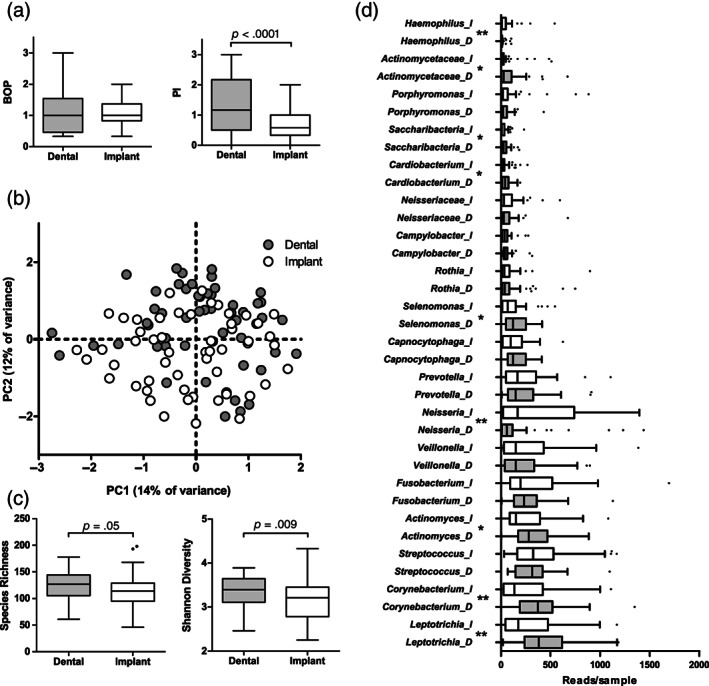
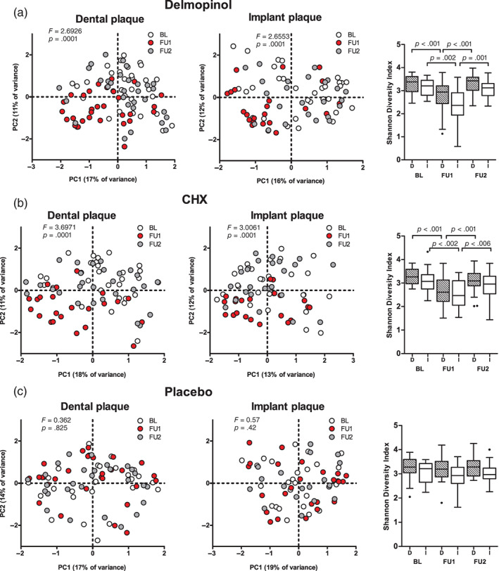
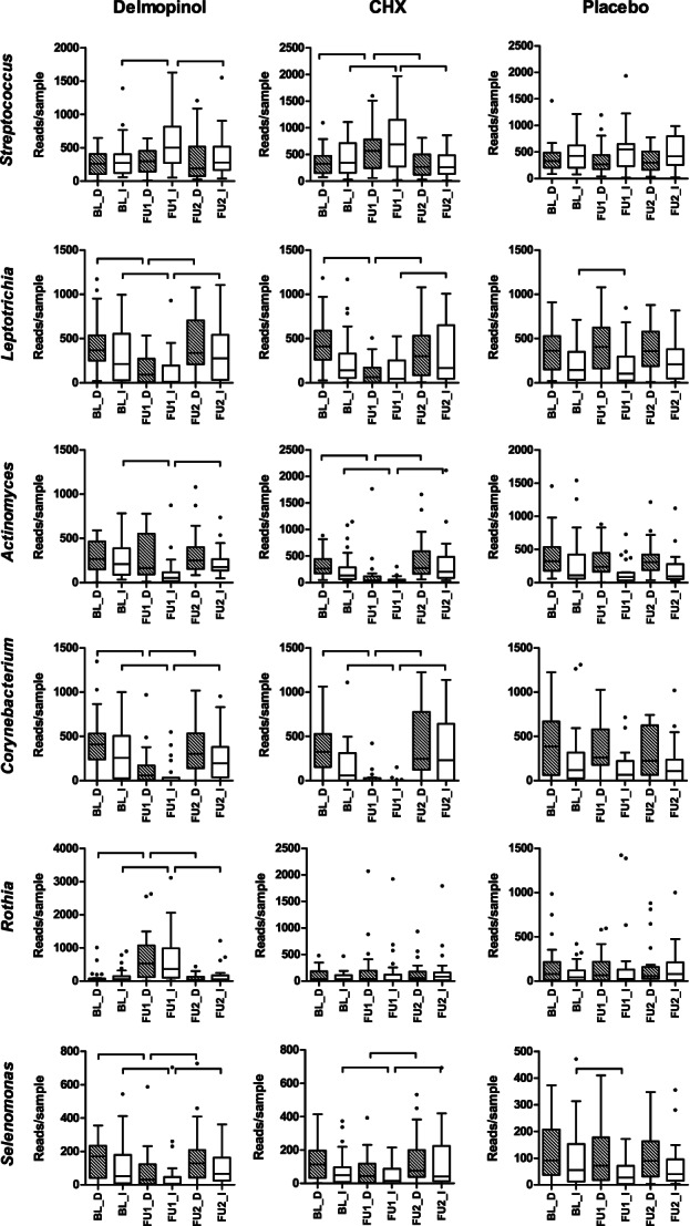
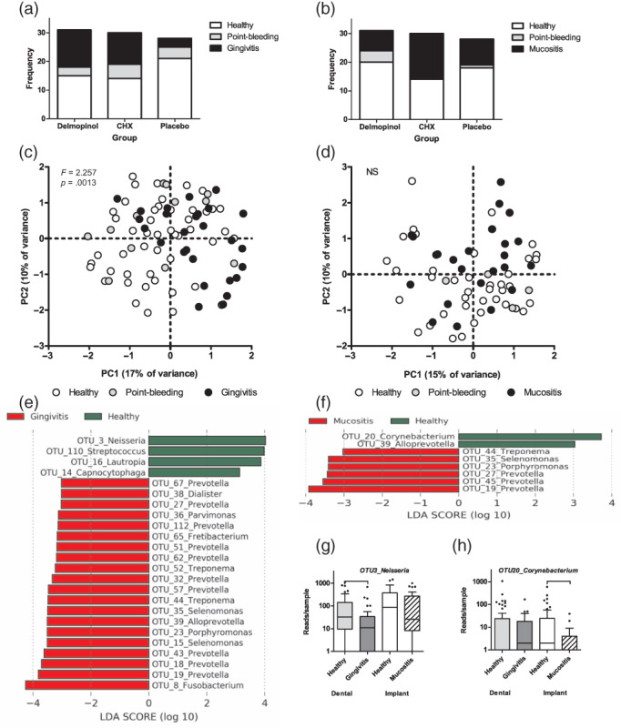
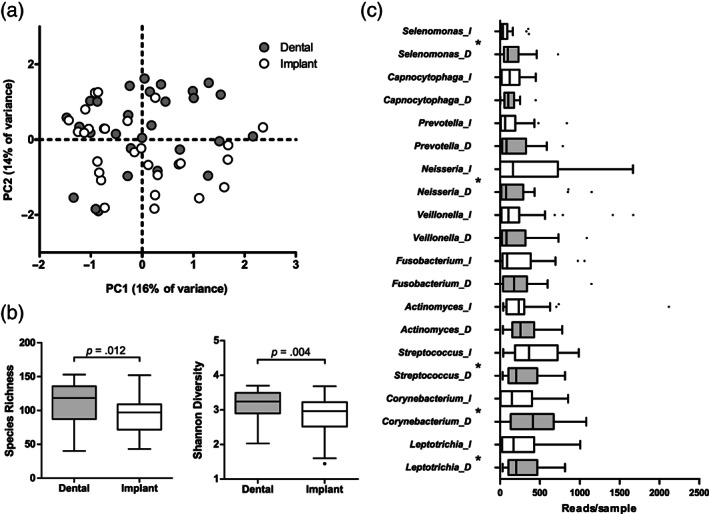
Results: The sites with peri-implant mucositis presented with a less diverse and less anaerobic microbiome. Exposure to delmopinol or CHX, but not to the placebo mouthrinse resulted in microbial changes after 1 month. The healthy sites around the teeth harboured a more diverse and more anaerobe-rich microbiome than the healthy sites around the implants.

Conclusions: Peri-implant sites with mucositis harbour ecologically less complex and less anaerobic biofilms with lower biomass than patient-matched dental sites with gingivitis while eliciting an equal inflammatory response. Adjunctive antimicrobial therapy in addition to mechanical debridement does affect both dental and peri-implant biofilm composition in the short term, resulting in a less dysbiotic subgingival biofilm.

Keywords: chlorhexidine; delmopinol; dental implant; microbiome; peri-implant mucositis; subgingival plaque; submucosal plaque.


Conflict of interest statement

The authors declare no conflict of interests.

Figures

FIGURE 1
FIGURE 1
Comparison of baseline subgingival plaque samples collected from paired dental sites with gingivitis and implant surfaces with mucositis. (a) Bleeding on probing (BOP) and plaque index (PI) around the sampled teeth and implants (p‐value based on Wilcoxon signed ranks test). (b) Principal component analysis (PCA) plot of the bacterial profile data (p = .0001, F = 2.84; restricted PERMANOVA). (c) Species richness (number of OTUs/sample) and Shannon diversity index (p‐values based on Wilcoxon signed ranks test) and (d) top 18 most abundant genera or higher taxa by sample type. D, samples from dental surfaces; I, samples from implant surfaces. Boxplots are based on Tukey. Statistical significance between the paired samples (Wilcoxon signed ranks test) at p < .05 is indicated with *, while p < .005 with **. p‐Values were not corrected for multiple comparisons. N = 54 dental and implant site pairs
FIGURE 2
FIGURE 2
Effects of 1‐month use of delmopinol (a), chlorhexidine (CHX); b), and placebo (c) mouthrinses on microbial profiles and diversity of subgingival plaque from dental surfaces and implant surfaces in time. BL, baseline visit; FU1, first follow‐up (1 month); FU2, second follow‐up (3 months). F‐ and p‐values are based on restricted PERMANOVA. Boxplots are based on Tukey. D, dental plaque; I, implant plaque. Connectors indicate a significant difference in diversity between timepoints (Wilcoxon signed ranks test) [Colour figure can be viewed at wileyonlinelibrary.com]
FIGURE 3
FIGURE 3
Examples of bacterial genera that were affected most significantly by the therapy. Hatched boxes indicate dental plaque and white boxes implant plaque. Connectors indicate a statistically significant difference (p < .05, Wilcoxon signed ranks test, p‐values not corrected for multiple comparisons). Full results on the 30 most abundant genera are shown in Table S3
FIGURE 4
FIGURE 4
Comparison of sampled sites 2 months after the therapy by diagnosis: frequency distribution by diagnosis per group at the dental sites (a) and the peri‐implant sites (b). Principal component analysis (PCA) plot of the bacterial profiles data of dental plaque (c) and peri‐implant plaque samples (d) by clinical diagnosis (F‐ and p‐values based on PERMANOVA, NS ‐ p > .05); the most significantly discriminating OTUs between healthy and gingivitis cases (e) and between healthy and mucositis cases (f) (here, LDA > 3; LEfSe; full list of discriminatory OTUs at LDA > 2 is presented in Table S2C). (g) Boxplots of OTU3_Neisseria, discriminating healthy and gingivitis samples. (h) Boxplots of OTU20_Corynebacterium, discriminating the healthy and mucositis samples. The two examples were the most discriminatory features from LEfSe output associated with soft tissue health [Colour figure can be viewed at wileyonlinelibrary.com]
FIGURE 5
FIGURE 5
Microbial profiles of healthy sites 2 months after the therapy (FU2) by sample type: (a) Principal component analysis (PCA) plot of the bacterial profile data of dental plaque and peri‐implant plaque (p = .033, F = 1.264, restricted PERMANOVA). (b) Species richness (number of OTUs/sample) and Shannon diversity index (p‐values based on Wilcoxon signed ranks test) and (c) top 10 most abundant genera by sample type. D, dental and I, peri‐implant plaque samples. Boxplots are based on Tukey. *Statistical significance between the paired samples at p < .05 (Wilcoxon signed ranks test). p‐Values were not corrected for multiple comparisons. N = 28 dental and implant sample pairs

References

    1. Belibasakis, G. N. (2014). Microbiological and immuno‐pathological aspects of peri‐implant diseases. Archives of Oral Biology, 59, 66–72. - PubMed
    1. Belibasakis, G. N. , & Manoil, D. (2021). Microbial community‐driven etiopathogenesis of peri‐implantitis. Journal of Dental Research, 100, 21–28. - PMC - PubMed
    1. Dabdoub, S. M. , Tsigarida, A. A. , & Kumar, P. S. (2013). Patient‐specific analysis of periodontal and peri‐implant microbiomes. Journal of Dental Research, 92, 168S–175S. - PMC - PubMed
    1. Daubert, D. , Pozhitkov, A. , McLean, J. , & Kotsakis, G. (2018). Titanium as a modifier of the peri‐implant microbiome structure. Clinical Implant Dentistry and Related Research, 20, 945–953. - PMC - PubMed
    1. Delgado‐Ruiz, R. , & Romanos, G. (2018). Potential causes of titanium particle and ion release in implant dentistry: A Systematic review. International Journal of Molecular Sciences, 19, 3585. - PMC - PubMed

Publication types

Substances