

2D morphometric analysis of Arabidopsis thaliana nuclei reveals characteristic profiles of different cell types and accessions
- PMID: 34665365
- PMCID: PMC8942920
- DOI: 10.1007/s10577-021-09673-2
2D morphometric analysis of Arabidopsis thaliana nuclei reveals characteristic profiles of different cell types and accessions
Erratum in
-
Correction to: 2D morphometric analysis of Arabidopsis thaliana nuclei reveals characteristic profiles of different cell types and accessions.Chromosome Res. 2022 Mar;30(1):25-27. doi: 10.1007/s10577-021-09677-y. Chromosome Res. 2022. PMID: 34962631 Free PMC article. No abstract available.
Abstract
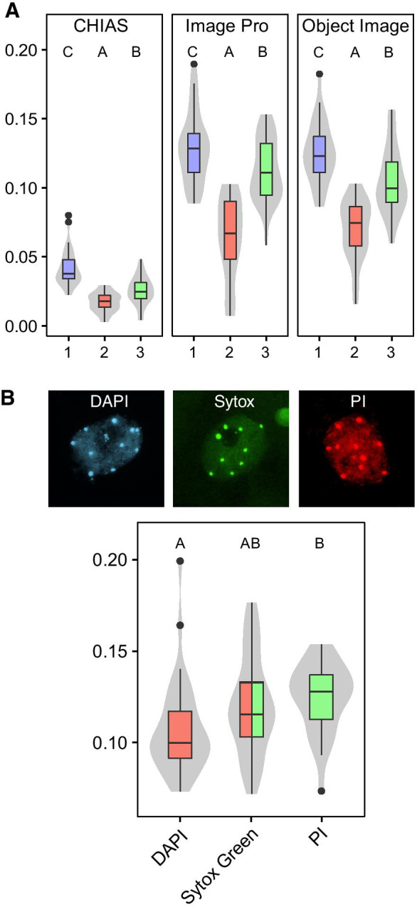
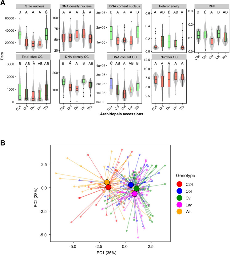
Functional changes of cells upon developmental switches and in response to environmental cues are often reflected in nuclear phenotypes, showing distinctive chromatin states corresponding to transcriptional changes. Such characteristic nuclear shapes have been microscopically monitored and can be quantified after differential staining of euchromatin and heterochromatin domains. Here, we examined several nuclear parameters (size, DNA content, DNA density, chromatin compaction, relative heterochromatin fraction (RHF), and number of chromocenters) in relation to spatial distribution of genes and transposon elements (TEs), using standard 2D fluorescence microscopy. We provide nuclear profiles for different cell types and different accessions of Arabidopsis thaliana. A variable, yet significant, fraction of TEs was found outside chromocenters in all cell types, except for guard cells. The latter cell type features nuclei with the highest level of chromatin compaction, while their chromocenters seem to contain gene-rich regions. The highest number of parameter correlations was found in the accession Cvi, whereas Ler showed only few correlations. This may point at differences in phenotype robustness between accessions. The significantly high association of NOR chromocenters in accessions Ws and Cvi corresponds to their low RHF level.
Keywords: Arabidopsis; Chromocenter; Heterochromatin; Nuclear phenotype; Quantitative analysis.
© 2021. The Author(s).
Conflict of interest statement
The authors declare no competing interests.
Figures


References
-
- Andrey P, Kiêu K, Kress C, Lehmann G, Tirichine L, Liu Z, Biot E, et al. Statistical analysis of 3D images detects regular spatial distributions of centromeres and chromocenters in animal and plant nuclei. PLoS Comput Biol. 2010;6(7):e1000853. doi: 10.1371/journal.pcbi.1000853.t002. - DOI - PMC - PubMed
-
- Arpòn A, Gaudin V, Andrey P (2018) A method for testing random spatial models on nuclear object distributions. In: Methods Mol Biol 2018;1675:493–507. doi: 10.1007/978-1-4939-7318-7_29 - PubMed
-
- Ashenafi MS, Baroux C (2018) Automated 3D gene position analysis using a customized Imaris Plugin: XTFISHInsideNucleus. Methods Mol Biol 1675:591–614. 10.1007/978-1-4939-7318-7_32 - PubMed
Publication types
MeSH terms
Substances
LinkOut - more resources
Full Text Sources
