. 2022 Jan;59(1):245-265.
doi: 10.1007/s12035-021-02511-w. Epub 2021 Oct 19.

Regulation of Brain Primary Cilia Length by MCH Signaling: Evidence from Pharmacological, Genetic, Optogenetic, and Chemogenic Manipulations

Affiliations

Regulation of Brain Primary Cilia Length by MCH Signaling: Evidence from Pharmacological, Genetic, Optogenetic, and Chemogenic Manipulations

Wedad Alhassen et al. Mol Neurobiol. 2022 Jan.

Abstract

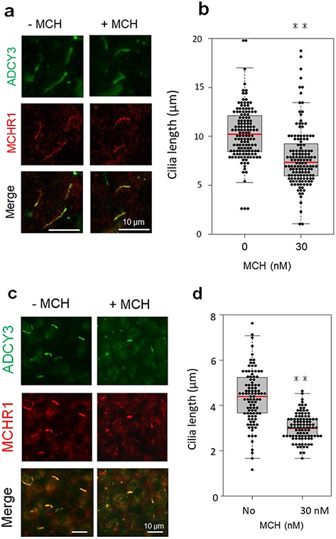
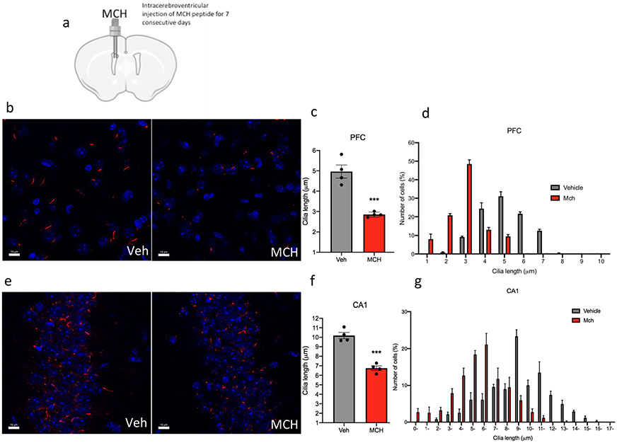
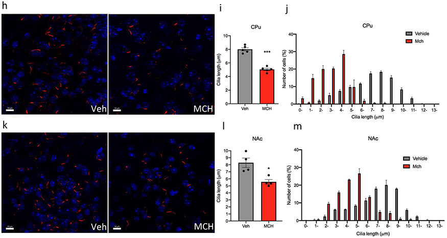
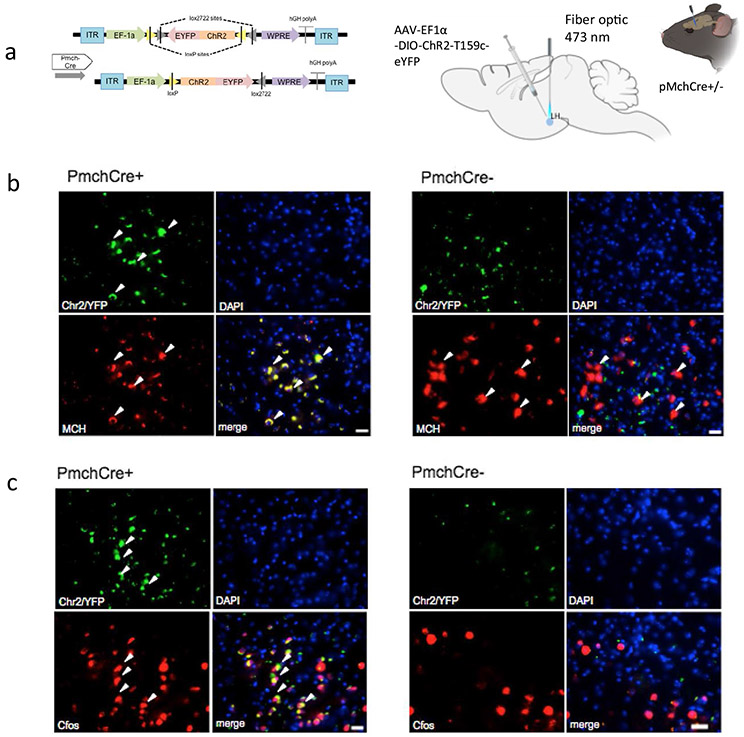
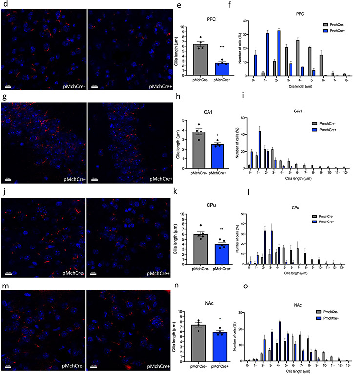
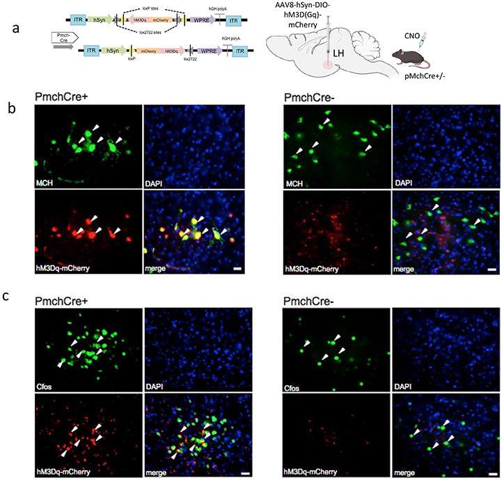
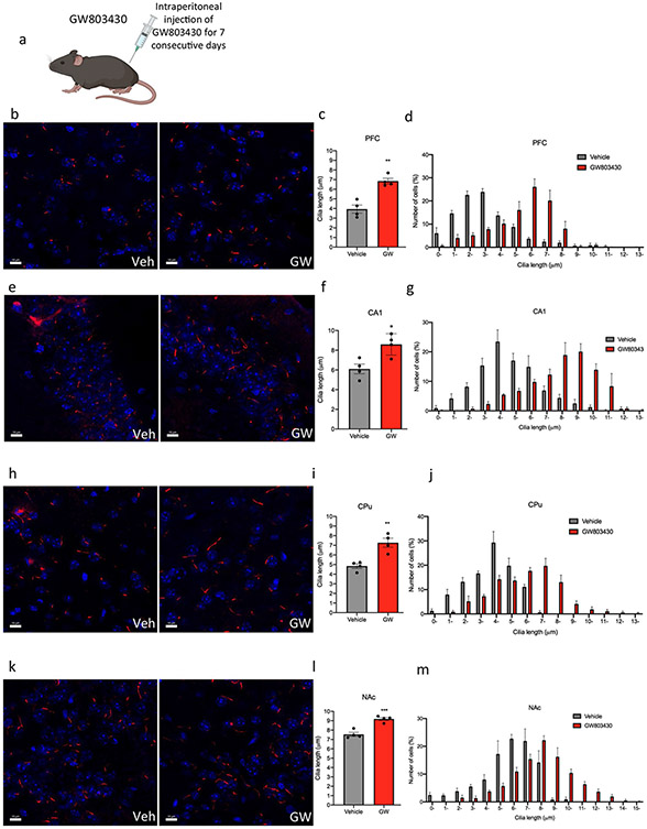
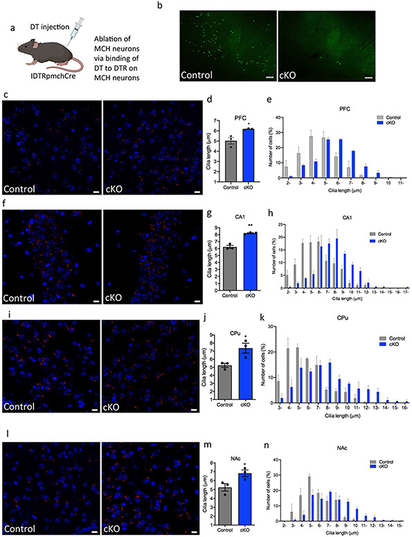
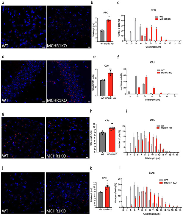
The melanin-concentrating hormone (MCH) system is involved in numerous functions, including energy homeostasis, food intake, sleep, stress, mood, aggression, reward, maternal behavior, social behavior, and cognition. In rodents, MCH acts on MCHR1, a G protein-coupled receptor, which is widely expressed in the brain and abundantly localized to neuronal primary cilia. Cilia act as cells' antennas and play crucial roles in cell signaling to detect and transduce external stimuli to regulate cell differentiation and migration. Cilia are highly dynamic in terms of their length and morphology; however, it is not known if cilia length is causally regulated by MCH system activation in vivo. In the current work, we examined the effects of activation and inactivation of MCH system on cilia lengths by using different experimental models and methodologies, including organotypic brain slice cultures from rat prefrontal cortex (PFC) and caudate-putamen (CPu), in vivo pharmacological (MCHR1 agonist and antagonist GW803430), germline and conditional genetic deletion of MCHR1 and MCH, optogenetic, and chemogenetic (designer receptors exclusively activated by designer drugs (DREADD)) approaches. We found that stimulation of MCH system either directly through MCHR1 activation or indirectly through optogenetic and chemogenetic-mediated excitation of MCH-neuron, caused cilia shortening, detected by the quantification of the presence of ADCY3 protein, a known primary cilia marker. In contrast, inactivation of MCH signaling through pharmacological MCHR1 blockade or through genetic manipulations - germline deletion of MCHR1 and conditional ablation of MCH neurons - induced cilia lengthening. Our study is the first to uncover the causal effects of the MCH system in the regulation of the length of brain neuronal primary cilia. These findings place MCH system at a unique position in the ciliary signaling in physiological and pathological conditions and implicate MCHR1 present at primary cilia as a potential therapeutic target for the treatment of pathological conditions characterized by impaired primary cilia function associated with the modification of its length.

Keywords: Activation; Brain; Cilia; Inactivation; Melanin-concentrating hormone; Signaling.


Figures

Fig. 1
Fig. 1
MCH treatment leads to cilia length shortening in cultured brain slices. CPu and PFC slices were treated with MCH on days 14 or 7 of culture, respectively. a, b Rat CPu slice cultures were treated with vehicle or 30 nM MCH for 18 h. c, d Rat PFC slice cultures were treated with vehicle or 30 nM MCH for 6 h. a, c Primary cilia were co-labeled with antibodies against ADCY3 (green) and MCHR1 (red). Scale bars = 10 μm. b, d The scatter plot represents cilium lengths measured using ADCY3/MCHR1 double labeling in randomly selected fields; at least 80 cilia per group in both CPu slices (b) and PFC slices (d) were evaluated, respectively. Primary cilia were significantly shorter in MCH-treated cultures than in control cultures for both CPu slices and PFC slices. Unpaired t test, **P < 0.01. Data are presented as means ± SEM
Fig. 2
Fig. 2
Chronic intracerebroventricular administration of MCH to mice shortens cilia length. a Schematic representation of chronic intracerebroventricular administration of MCH peptide. Male mice were administered with vehicle or MCH (1 nmol). Diagram was created with BioRender.com webpage. b, e, h, k Immunostaining of ADCY3 labeled in red fluorescence and counterstained with DAPI (blue) in the b PFC, e CA1, h CPu, and k NAc in animals administered with MCH or vehicle (scale bar = 10 μm, magnification = 63 ×). c, e, g, i Quantification of the length of ADCY3 + primary cilia (μm) in the c PFC, e CA1, g CPu, and (i) NAc in animals administered with MCH or vehicle, ***P < 0.0007 and **P < 0.01. d, g, j, m Cilia were grouped by length (μm) and plotted against the number of cells (%) in the d PFC, g CA1, j CPu, and m NAc
Fig. 2
Fig. 2
Chronic intracerebroventricular administration of MCH to mice shortens cilia length. a Schematic representation of chronic intracerebroventricular administration of MCH peptide. Male mice were administered with vehicle or MCH (1 nmol). Diagram was created with BioRender.com webpage. b, e, h, k Immunostaining of ADCY3 labeled in red fluorescence and counterstained with DAPI (blue) in the b PFC, e CA1, h CPu, and k NAc in animals administered with MCH or vehicle (scale bar = 10 μm, magnification = 63 ×). c, e, g, i Quantification of the length of ADCY3 + primary cilia (μm) in the c PFC, e CA1, g CPu, and (i) NAc in animals administered with MCH or vehicle, ***P < 0.0007 and **P < 0.01. d, g, j, m Cilia were grouped by length (μm) and plotted against the number of cells (%) in the d PFC, g CA1, j CPu, and m NAc
Fig. 3
Fig. 3
Optogenetic stimulation of MCH shortens cilia length. a Experimental approach. Adult PmchCre + and PmchCre− mice were stereotaxically injected with AAV-EF1α-DIO-ChR2-T159c-eYFP in the lateral hypothalamus (LH). A fiber cannula was then placed in the LH slightly above the injection site. Diagram was created with BioRender.com webpage. b Coexpression of ChR2 (green) containing neurons in the lateral hypothalamus and MCH immunofluorescence (red) and counterstained with DAPI (blue) in PmchCre + and PmchCre− mice. (scale bar = 10 μm, magnification = 40×). c ChR2 fluorescence (green) and c-Fos immunofluorescence (red) identifies cells recently activated in PmchCre + and PmchCre− mice (scale bar = 10 μm). d, g, j, m Immunostaining of ADCY3 labeled in red fluorescence in the d PFC, g CA1, j CPu, and m NAc after blue light activation in PmchCre + and PmchCre− mice (scale bar = 10 μm). e, h, k, n Quantification of the length of ADCY3 + primary cilia (μm) in the e PFC, h CA1, k CPu, and n NAc, *P < 0.05, **P < 0.01, and ***P < 0.001. f, I, l, o Cilia were grouped by length (μm) and plotted against the number of cells (%) in the f PFC, i CA1, l CPu, and o NAc
Fig. 3
Fig. 3
Optogenetic stimulation of MCH shortens cilia length. a Experimental approach. Adult PmchCre + and PmchCre− mice were stereotaxically injected with AAV-EF1α-DIO-ChR2-T159c-eYFP in the lateral hypothalamus (LH). A fiber cannula was then placed in the LH slightly above the injection site. Diagram was created with BioRender.com webpage. b Coexpression of ChR2 (green) containing neurons in the lateral hypothalamus and MCH immunofluorescence (red) and counterstained with DAPI (blue) in PmchCre + and PmchCre− mice. (scale bar = 10 μm, magnification = 40×). c ChR2 fluorescence (green) and c-Fos immunofluorescence (red) identifies cells recently activated in PmchCre + and PmchCre− mice (scale bar = 10 μm). d, g, j, m Immunostaining of ADCY3 labeled in red fluorescence in the d PFC, g CA1, j CPu, and m NAc after blue light activation in PmchCre + and PmchCre− mice (scale bar = 10 μm). e, h, k, n Quantification of the length of ADCY3 + primary cilia (μm) in the e PFC, h CA1, k CPu, and n NAc, *P < 0.05, **P < 0.01, and ***P < 0.001. f, I, l, o Cilia were grouped by length (μm) and plotted against the number of cells (%) in the f PFC, i CA1, l CPu, and o NAc
Fig. 4
Fig. 4
Chemogenic excitation of MCH shortens cilia length. a Experimental approach. Adult PmchCre + and PmchCre− mice underwent bilateral stereotaxic injection in the lateral hypothalamus with AAV-hSyn-DIO-hM3D(Gq)-mCherry to express DREADD in Cre-expressing MCH neurons. Clozapine-N-oxide (CNO) was delivered intraperitoneally to stimulate MCH neurons. Diagram was created with BioRender.com webpage. b Co-expression of mCherry fluorescence (red) identifying DREADD-expressing neurons and MCH immunofluorescence (green) and counterstained with DAPI (blue) in Pmch-Cre+and PmchCre− mice (scale bar = 10 μm, magnification = 40 ×). c mCherry fluorescence (red) identifies DREADD-expressing neurons, and c-Fos immunofluorescence (green) identifies cells recently activated in PmchCre + and PmchCre− mice (Scale bar = 10 μm). d, g, j, m Immunostaining of ADCY3 labeled in red fluorescence in the d PFC, g CA1, j CPu and m NAc via CNO-dependent activation of Gq signaling in PmchCre + and PmchCre− mice (scale bar = 10 μM). e, h, k, n) Quantification of the length of ADCY3 + primary cilia in the e PFC, h CA1, k CPu, and n NAc, **P < 0.01 and ***P < 0.001. f, i, l, o Cilia were grouped by length (μm) and plotted against the number of cells (%) in the f PFC, i CA1, l CPu, and o NAc
Fig. 4
Fig. 4
Chemogenic excitation of MCH shortens cilia length. a Experimental approach. Adult PmchCre + and PmchCre− mice underwent bilateral stereotaxic injection in the lateral hypothalamus with AAV-hSyn-DIO-hM3D(Gq)-mCherry to express DREADD in Cre-expressing MCH neurons. Clozapine-N-oxide (CNO) was delivered intraperitoneally to stimulate MCH neurons. Diagram was created with BioRender.com webpage. b Co-expression of mCherry fluorescence (red) identifying DREADD-expressing neurons and MCH immunofluorescence (green) and counterstained with DAPI (blue) in Pmch-Cre+and PmchCre− mice (scale bar = 10 μm, magnification = 40 ×). c mCherry fluorescence (red) identifies DREADD-expressing neurons, and c-Fos immunofluorescence (green) identifies cells recently activated in PmchCre + and PmchCre− mice (Scale bar = 10 μm). d, g, j, m Immunostaining of ADCY3 labeled in red fluorescence in the d PFC, g CA1, j CPu and m NAc via CNO-dependent activation of Gq signaling in PmchCre + and PmchCre− mice (scale bar = 10 μM). e, h, k, n) Quantification of the length of ADCY3 + primary cilia in the e PFC, h CA1, k CPu, and n NAc, **P < 0.01 and ***P < 0.001. f, i, l, o Cilia were grouped by length (μm) and plotted against the number of cells (%) in the f PFC, i CA1, l CPu, and o NAc
Fig. 5
Fig. 5
Systemic administration of the GW803430 (MCHR1 antagonist) to mice increases cilia length. a Schematic representation of systemic administration of GW803430 or vehicle for 7 consecutive days in adult mice. Diagram was created with BioRender.com webpage. b, e, h, k Immunostaining of ADCY3 labeled in red fluorescence in the b PFC, e CA1, h CPu, and k NAc in animals administered with GW803430 or vehicle (scale bar = 10 μm). c, f, I, l Quantification of the length of ADCY3 + primary cilia in the c PFC, f CA1, i CPu, and l NAc in animals administered with GW803430 or vehicle, *P < 0.05, **P < 0.01, and ***P < 0.001. d, g, j, m Cilia were grouped by length (μm) and plotted against the number of cells (%) in the d PFC, g CA1, j CPu, and m NAc
Fig. 6
Fig. 6
Cilia length is increased in MCH deficit mice. a Schematic representation of how the MCH cells are ablated through DTR. IDTR mice were crossed with PmchCre mice rendering offspring mice with Cre-expressing MCH neurons sensitive to DT. Diagram was created with BioRender.com webpage. b MCH immunoreactivity (GFP) in the lateral hypothalamus of IDTRPmchCre− and IDTRPmchCre + mice following DT injection (scale bar = 100 μm, magnification = 10 ×). c, f, I, l Immunostaining of ADCY3 labeled in red fluorescence and counterstained with DAPI (blue) in IDTRPmch + and IDTRPmch− mice in the c PFC, e CA1, g CPu, and i NAc (scale bar = 10 μm, magnification = 63 ×). d, g, j, m Quantification of cilia length (μm) in the d PFC, f CA1, h CPu, and j NAc, *P < 0.05 and **P < 0.01. e, h, k, n Cilia were grouped by length (μm) and plotted against the number of cells (%) in the e PFC, h CA1, k CPu, and n NAc
Fig. 7
Fig. 7
Cilia length is increased in MCHR1 knockout animals. Immunostaining of ADCY3 labeled in red fluorescence and counterstained with DAPI (blue) in the a PFC, d CA1, g CPu, and j NAc in MCHR1 KO or WT mice (scale bar = 10 μm, magnification = 63 ×. b, e, h, k Quantification of the length of ADCY3+ primary cilia (μm) in the b PFC, e CA1, h CPu (CPu), and k NAc in MCHR1 KO or WT mice, **P < 0.01 and ***P < 0.001. c, f, I, l Cilia were grouped by length (μm) and plotted against the number of cells (%) in the c PFC, f CA1, i CPu, and l NAc

References

    1. Alachkar A, Alhassen L, Wang Z, Wang L, Onouye K, Sanathara N, Civelli O (2016) Inactivation of the melanin concentrating hormone system impairs maternal behavior. European neuropsychopharmacology: the journal of the European College of Neuropsychopharmacology 26:1826–1835 - PMC - PubMed
    1. Alhassen L, Phan A, Alhassen W, Nguyen P, Lo A, Shaharuddin H, Sanathara N, Civelli O, Alachkar A (2019) The role of olfaction in MCH-regulated spontaneous maternal responses. Brain Res 1719:71–76 - PubMed
    1. Alhassen W, Chen S, Vawter M, Robbins BK, Nguyen H, Myint TN, Saito Y, Schulmann A, Nauli SM, Civelli O et al. (2021) Patterns of cilia gene dysregulations in major psychiatric disorders. Prog Neuropsychopharmacol Biol Psychiatry 109:110255. - PMC - PubMed
    1. Arletti R, Benelli A, Bertolini A (1992) Oxytocin involvement in male and female sexual behavior. Ann N Y Acad Sci 652:180–193 - PubMed
    1. Atkinson KF, Sherpa RT, and Nauli SM (2019). The role of the primary cilium in sensing extracellular pH. Cells 8.

MeSH terms

Substances