. 2021 Apr;184(4):607-615.
doi: 10.1530/EJE-20-1268.

The natural history of 21-hydroxylase autoantibodies in autoimmune Addison's disease

Affiliations

The natural history of 21-hydroxylase autoantibodies in autoimmune Addison's disease

Anette Boe Wolff et al. Eur J Endocrinol. 2021 Apr.

Abstract

Background: The most common cause of primary adrenal failure (Addison's disease) in the Western world is autoimmunity characterized by autoantibodies against the steroidogenic enzyme 21-hydroxylase (CYP21A2, 21OH). Detection of 21OH-autoantibodies is currently used for aetiological diagnosis, but how levels of 21OH-autoantibodies vary over time is not known.

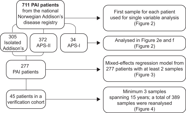
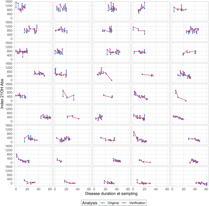
Setting: Samples from the national Norwegian Addison's Registry and Biobank established in 1996 (n = 711). Multi-parameter modelling of the course of 21OH-autoantibody indices over time.

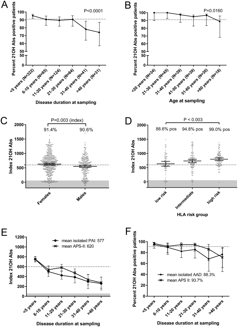
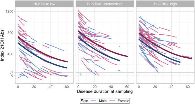
Results: 21OH-autoantibody positivity is remarkably stable, and >90% of the patients are still positive 30 years after diagnosis. Even though the antibody levels decline with disease duration, it is only rarely that this downturn reaches negativity. 21OH-autoantibody indices are affected by age at diagnosis, sex, type of Addison's disease (isolated vs autoimmune polyendocrine syndrome type I or II) and HLA genotype.

Conclusion: 21OH-autoantibodies are reliable and robust markers for autoimmune Addison's disease, linked to HLA risk genotype. However, a negative test in patients with long disease duration does not exclude autoimmune aetiology.


Figures

Figure 1
Figure 1
Flow chart describing the patients in this study.
Figure 2
Figure 2
Individual variables’ impact on 21OH-Abs frequency and indices (A) Percentage of isolated PAI and APS-II patients positive for antibodies against 21OH (21OH-Abs) separated in groups according to disease duration at sampling. The statistical test was done with logistic regression analysis. (B) Percentage of isolated PAI and APS-II patients positive for 21OH-Abs sampled within 1 year of diagnosis in relation to age at diagnosis. Statistical analysis was done with logistic regression analysis. (C) 21OH-Abs-indicies in males and females. Percentage positive for each sex is shown inside the corresponding box. Statistical test for indices between the groups was achieved by a parametric t-test while statistical differences between the frequencies were done by a Pearson’s chi test. (D) 21OH-Abs indices stratified by HLA-risk groups given in Table 2 and percentage positives in each group. Statistical differences were calculated by ANOVA and Tukey’s test. (E) 21OH-Abs-indices stratified into phenotypic groups and disease duration at sampling; isolated PAI (black) and APS-II (red). (F) Percent positive for 21OH-Abs stratified to disease duration at sampling; isolated PAI (black) and APS-II (red). The dashed line (A, B and F) represents the mean positivity for autoantibodies against 21OH-Abs in the whole isolated, AAD autoimmune PAI and APS-II cohort. The dashed line (C and E) represents the mean indices of 21OH-Abs. The shaded area (C–E) in the lower part of the graph represent where the test gives negative results (sets the threshold for positivity). 95% CI is shown by vertical bars.
Figure 3
Figure 3
Estimated individual trajectories from mixed-effects longitudinal regression model for 21OH-Abs (n = 1287 observations from 277 individuals). The lines corresponds to individual patients. Includes data from patients with at least two samples. The thick lines show the predicted trajectory for a ‘typical’ patient (all random effects set to 0), that is a 40-year-old female with APS-II.
Figure 4
Figure 4
Course of 21OH-Abs in the 45 patients from the verification study (n = 389 samples) assayed at inclusion in the registry and in the verification study.

References

    1. Husebye ES, Allolio B, Arlt W, Badenhoop K, Bensing S, Betterle C, Falorni A, Gan EH, Hulting AL, Kasperlik-Zaluska Aet al. Consensus statement on the diagnosis, treatment and follow-up of patients with primary adrenal insufficiency. Journal of Internal Medicine 2014. 275 104–115. (10.1111/joim.12162) - DOI - PubMed
    1. Saverino S, Falorni A.Autoimmune Addison's disease. Best Practice and Research: Clinical Endocrinology and Metabolism 2020. 34 101379. (10.1016/j.beem.2020.101379) - DOI - PubMed
    1. Betterle C, Presotto F, Furmaniak J.Epidemiology, pathogenesis, and diagnosis of Addison's disease in adults. Journal of Endocrinological Investigation 2019. 42 1407–1433. (10.1007/s40618-019-01079-6) - DOI - PubMed
    1. Conrad K, Roggenbuck D, Reinhold D, Sack U.Autoantibody diagnostics in clinical practice. Autoimmunity Reviews 2012. 11 207–211. (10.1016/j.autrev.2011.05.014) - DOI - PubMed
    1. Erichsen MM, Lovas K, Fougner KJ, Svartberg J, Hauge ER, Bollerslev J, Berg JP, Mella B, Husebye ES.Normal overall mortality rate in Addison's disease, but young patients are at risk of premature death. European Journal of Endocrinology 2009. 160 233–237. (10.1530/EJE-08-0550) - DOI - PubMed