. 2021 Nov 12;18(6):10.1088/1741-2552/ac310a.
doi: 10.1088/1741-2552/ac310a.

Light-weight electrophysiology hardware and software platform for cloud-based neural recording experiments

Affiliations

Light-weight electrophysiology hardware and software platform for cloud-based neural recording experiments

Kateryna Voitiuk et al. J Neural Eng. .

Abstract

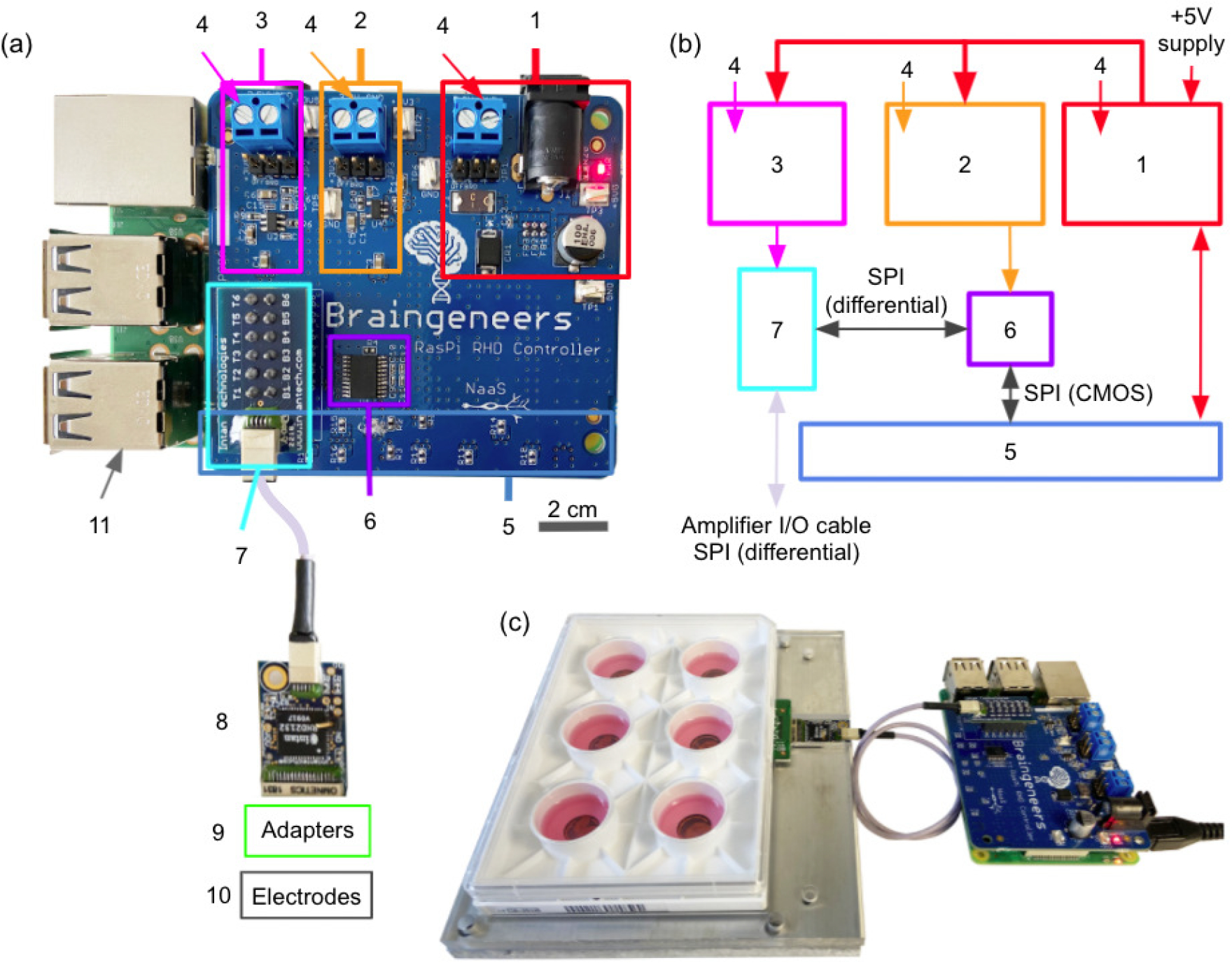
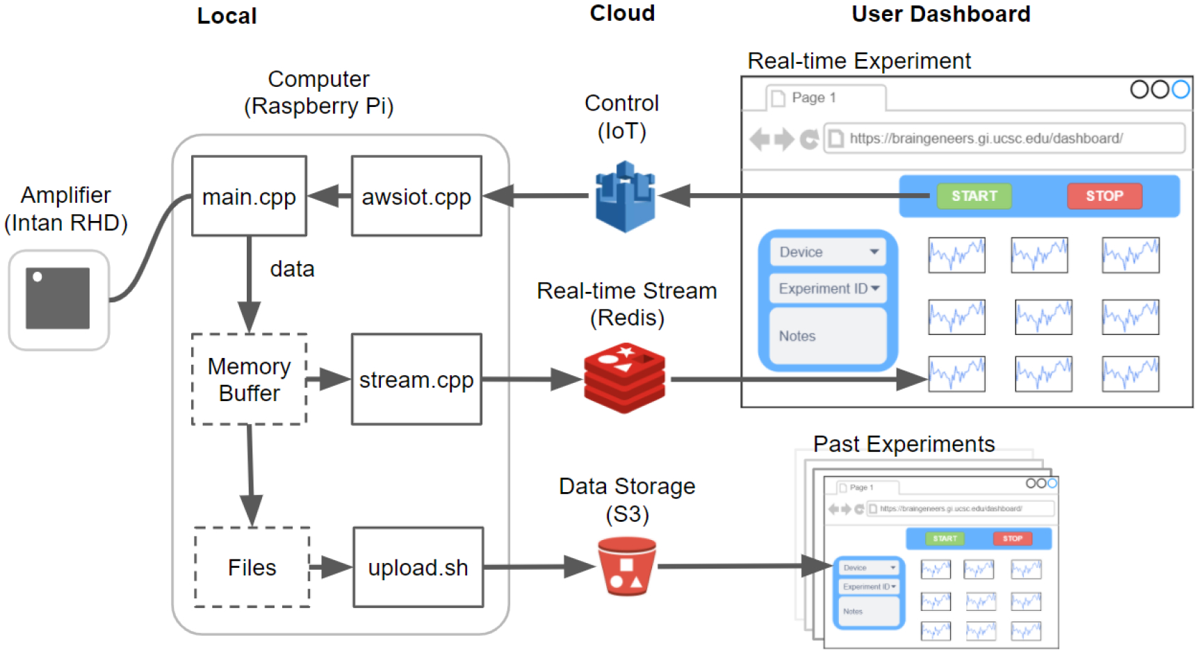
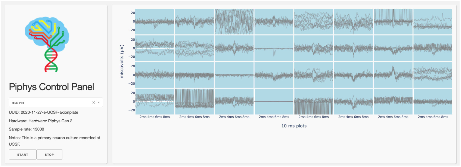
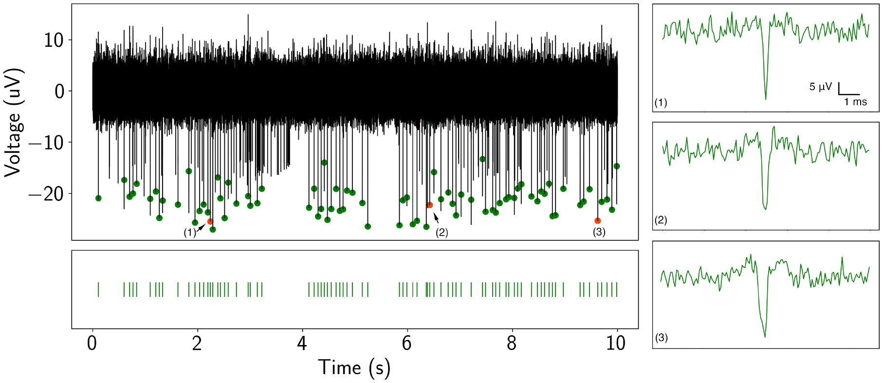
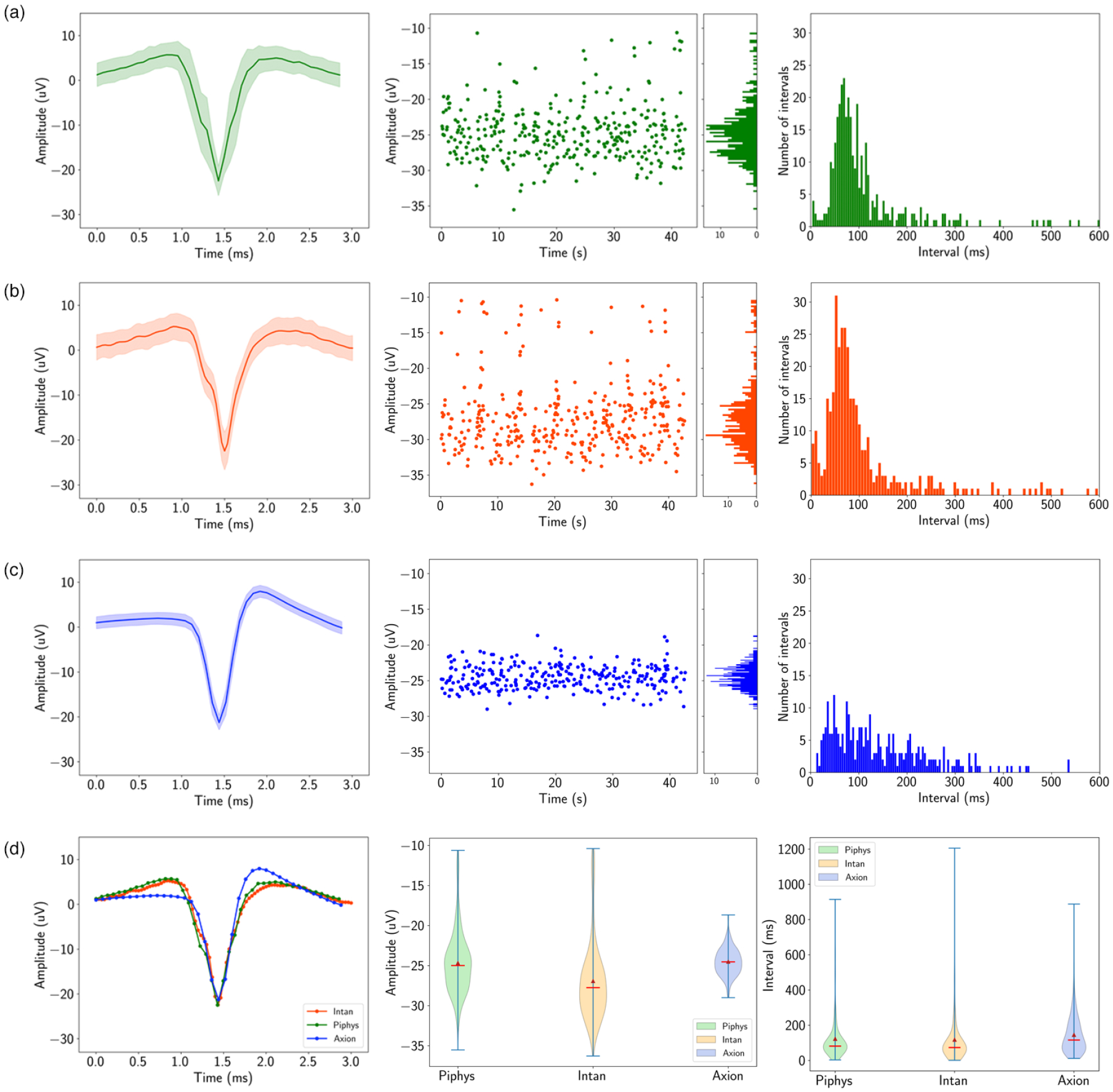
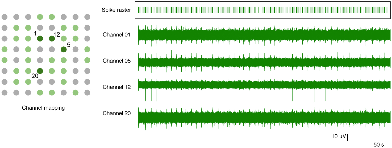
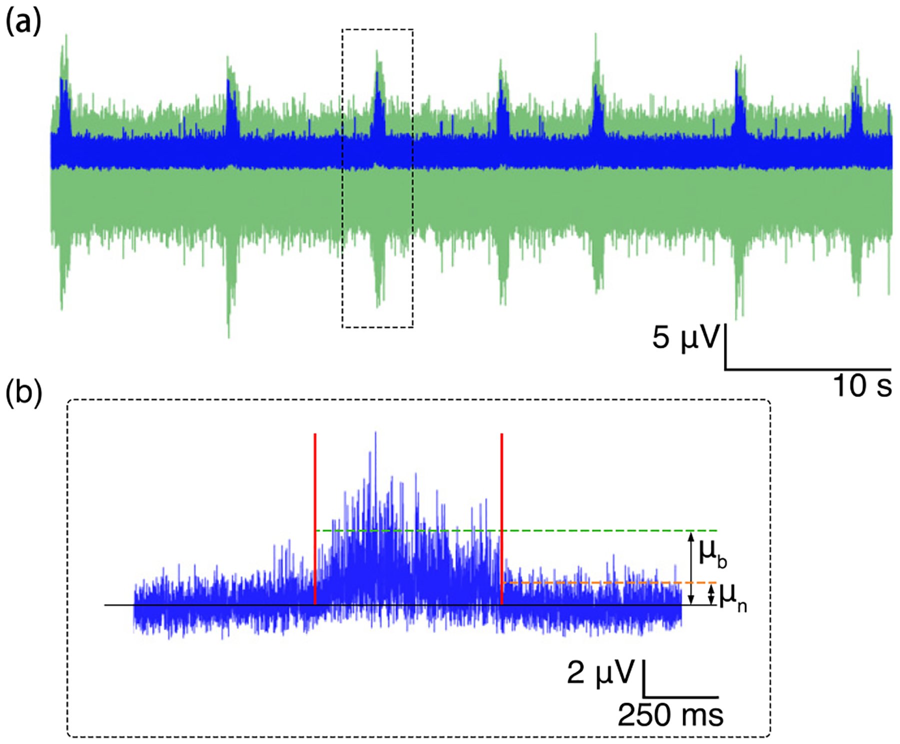
Objective.Neural activity represents a functional readout of neurons that is increasingly important to monitor in a wide range of experiments. Extracellular recordings have emerged as a powerful technique for measuring neural activity because these methods do not lead to the destruction or degradation of the cells being measured. Current approaches to electrophysiology have a low throughput of experiments due to manual supervision and expensive equipment. This bottleneck limits broader inferences that can be achieved with numerous long-term recorded samples.Approach.We developed Piphys, an inexpensive open source neurophysiological recording platform that consists of both hardware and software. It is easily accessed and controlled via a standard web interface through Internet of Things (IoT) protocols.Main results.We used a Raspberry Pi as the primary processing device along with an Intan bioamplifier. We designed a hardware expansion circuit board and software to enable voltage sampling and user interaction. This standalone system was validated with primary human neurons, showing reliability in collecting neural activity in near real-time.Significance.The hardware modules and cloud software allow for remote control of neural recording experiments as well as horizontal scalability, enabling long-term observations of development, organization, and neural activity at scale.

Keywords: IoT; data acquisition; electrophysiology; in vitro; neural recording; open source; scalable.


Conflict of interest statement

Competing interests

The authors declare no conflict of interest.

Figures

Figure 1.
Figure 1.
Cloud-based experiment paradigm: biological measurement and local hardware are presented to the user through the cloud, such that experiment management and control can be administrated remotely and may be automated by a computer program.
Figure 2.
Figure 2.. Piphys hardware components.
(a) Expansion shield (blue board) attached on top of Raspberry Pi (green board). (b) Logic level connection. (c) Example interface with standard 6-well electrode plate. (1) +5V logic, (2) +3.3V logic, (3) +3.5V logic, (4) External supply inputs, (5) Raspberry Pi input/output pins (bottom), (6) LVDS converter, (7) Intan RHD adapter, (8) Intan RHD 32-channel recording headstage containing Intan RHD2132 bioamplifier chip, (9) Optional adapter board to electrodes, (10) Multiple electrode types possible, (11) Raspberry Pi computer (bottom).
Figure 3.
Figure 3.. Software overview.
The software that runs on the local Raspberry Pi device communicates with the Intan RHD2132 bioamplifier chip to stream and store the digitized neural signal. Concurrently, it pushes the signal to Redis for near real-time visualization on the online dashboard. Datasets are also uploaded to S3 after each recording for permanent storage and access. Experimental control such as ‘start’, ‘stop’, and variable configuration is sent from the dashboard through Amazon IoT to the local device. Past experiment data can also be browsed using records from S3.
Figure 4.
Figure 4.. Dashboard.
A control panel interface is displayed through the browser running spike detection by thresholding.
Figure 5.
Figure 5.. Detection of neuronal spike activity using Piphys.
Spike train (black trace) from a recorded neuron in the time domain from Piphys. Spikes shown here are sorted from SpyKING CIRCUS software and labeled on the raw data with green and orange dots. Bottom: spike raster is aligned with the detected spikes showing firing activities at specific positions. (1)(2)(3) Individual spike examples randomly picked from the spike train. The analysis and quantification of the spike train and waveform are shown in Figure 6.
Figure 6.
Figure 6.. Piphys performance is similar to commercial systems.
Spike sorting result for the same recording channel from Piphys, Intan RHD USB interface board, and Axion Maestro Edge. Shown from left to right are mean waveform with standard deviation (shaded area), amplitudes of the detected spikes over time, and interspike interval distribution. (Figure 5 shows a small sample of one of the channels measured using the Piphys system) (a) Piphys has −24.67 ±3.92μV for the mean waveform, firing rate of 8.05 spikes/s, and mean interspike interval of 122.79 ms. (b) Intan RHD USB interface board has −26.92 ±4.96μV for the mean waveform, firing rate of 8.44 spikes/s, and mean interspike interval of 118.15 ms. (c) Axion Maestro Edge has −24.50 ±1.69μV for the mean waveform, firing rate of 6.86 spikes/s, and mean interspike interval of 145.57 ms. (d) Comparison of the mean waveform, amplitude, and interspike interval distribution from three systems.
Figure 7.
Figure 7.. Bursting activity across four channels with channel mapping.
Channel mapping shows 64 electrodes in well B2 of the Axion plate. Light green dots are the 32 electrodes recorded by Piphys. Dark green dots mark channels 1, 5, 12 and 20 whose raw recording plots are on the right. The spike raster superimposes all the detected spikes in the shown channels. Each light green vertical line in the raster indicates a spike, and the dark green bar is the result of superimposing multiple spikes in the burst. The bars in the raster plot align with the bursts throughout these four channels. Burst population rate is 0.13 per second. The number of spikes in each burst is 55 ± 17.58.
Figure 8.
Figure 8.. Signal-to-noise ratio of the burst.
(a) Burst train (green) and the smoothed signal (blue). (b) Zoom in to the third smoothed burst showing means of the signal and the baseline noise for SNR calculation.

References

    1. Hansel D, Mato G, and Meunier C. Synchrony in Excitatory Neural Networks. Neural Computation, 7(2):307–337, March 1995. Publisher: MIT Press. - PubMed
    1. Beggs John M. and Plenz Dietmar. Neuronal Avalanches in Neocortical Circuits. Journal of Neuroscience, 23(35):11167–11177, December 2003. Publisher: Society for Neuroscience Section: Behavioral/Systems/Cognitive. - PMC - PubMed
    1. Poli Daniele, Pastore Vito P., and Massobrio Paolo. Functional connectivity in in vitro neuronal assemblies. Frontiers in Neural Circuits, 9, 2015. Publisher: Frontiers. - PMC - PubMed
    1. Huang Yu-Ting, Chang Yu-Lin, Chen Chun-Chung, Lai Pik-Yin, and Chan CK. Positive feedback and synchronized bursts in neuronal cultures. PLOS ONE, 12(11):e0187276, November 2017. Publisher: Public Library of Science. - PMC - PubMed
    1. Trujillo Cleber A., Gao Richard, Negraes Priscilla D., Gu Jing, Buchanan Justin, Preissl Sebastian, Wang Allen, Wu Wei, Haddad Gabriel G., Chaim Isaac A., Domissy Alain, Vandenberghe Matthieu, Devor Anna, Yeo Gene W., Voytek Bradley, and Muotri Alysson R.. Complex Oscillatory Waves Emerging from Cortical Organoids Model Early Human Brain Network Development. Cell Stem Cell, 25(4):558–569.e7, October 2019. - PMC - PubMed

Publication types