. 2021 Oct 19;7(1):63.
doi: 10.1186/s40942-021-00333-5.

Lamellar Hole-associated Epiretinal Proliferation in choroideremia: a case report

Affiliations

Lamellar Hole-associated Epiretinal Proliferation in choroideremia: a case report

Vittoria Murro et al. Int J Retina Vitreous. .

Abstract

Background: To report a clinical case of a patient affected with choroideremia (CHM) who underwent macular surgery for a macular hole (MH) with Lamellar Hole-associated Epiretinal Proliferation (LHEP).

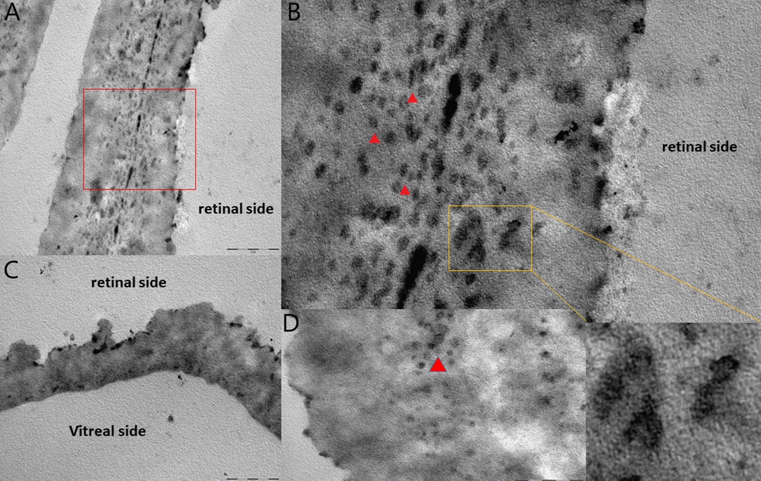
Case presentation: We have described a 48-year-old male patient affected with CHM who developed MH with LHEP over a 7-year follow-up. The patient was referred to the Regional Center for Hereditary Retinal Degenerations of the Eye Clinic in Florence (Italy) in April 2012. The patient underwent vitrectomy and Inner Limiting Membrane (ILM) and LHEP peeling with fluid-air exchange. Ultra-structural examination of the excised epiretinal proliferation, carried out using electron microscopy, showed dense amorphous material, mainly composed of abundant clusters of fibrous collagens resembling compact fibrous long spacing collagen (FLSC), embedded in native vitreous collagen (NVC) and type IV collagen. No cells were detected in any of the specimens collected. At the 3rd-week postoperative follow-up the macular hole was closed.

Conclusion: Macular hole with LHEP can be detected in CHM patients; in our patient the macular hole showed tractional and degenerative features, with good anatomical results after macular surgery.

Keywords: CHM; Electron microscopy; LHEP; Lamellar Hole-associated Epiretinal Proliferation; Macular hole; OCT; Peeling; Vitrectomy.


Conflict of interest statement

The authors declare that they have no competing interests.

Figures

Fig. 1
Fig. 1
(A, B) Color fundus photographs (CFP) of the RE and LE respectively showing extensive chorioretinal atrophy at the referral in April 2012 (A) and in May 2017 (B). (C) Fundus autofluorescence (Blue-FAF) imaging showing an island of preserved RPE in the macula of both eyes. (D, E) OCT examination of the RE in 2012 (D) and in May 2017 (E) showing very thin neuroepithelium. (F) In October 2018, Epiretinal Proliferation (LHEP) is clearly visible attached to the inner surface of the retina. The red rectangle shows the magnified area in (G); the LHEP is outlined by yellow arrowheads. Two intraretinal cysts (blue asterisks) are visible at the inner nuclear layer and fine hyper-reflective bridges of tissue (blue arrows) across the neurosensory retina. Outer retinal tubulations (red circle) are detectable. (H) Post-surgical OCT scan shows closed macular hole (January 2019). (I) CFP of the RE and LE in July 2020; (J) OCT examination of the RE in July 2020. (K) Infrared-reflectance and OCT imaging of the left eye
Fig. 2
Fig. 2
AD Transmission electron microscopy images of epiretinal tissue. Image A showing dense extracellular matrix constituted of amorphous material of collagen strands (native vitreous collagen NVC and type IV collagen). The red square shows the magnified area in B. In Image B abundant clusters of fibrous collagen closely resembling compact FLSC (red arrowheads) embedded in native vitreous collagen (NVC) and type IV collagen are detectable. C The vitreal side of the epiretinal tissue appear smoother than the retinal side. D FLSC (detail in yellow square) embedded in vitreous collagen fibrils

References

    1. Mitsios A, Dubis AM, Moosajee M. Choroideremia: from genetic and clinical phenotyping to gene therapy and future treatments. Ther Adv Ophthalmol. 2018;10:2515841418817490. - PMC - PubMed
    1. Genead MA, Fishman GA. Cystic macular oedema on spectral-domain optical coherence tomography in choroideremia patients without cystic changes on fundus examination. Eye (Lond) 2010;25(1):84–90. doi: 10.1038/eye.2010.157. - DOI - PMC - PubMed
    1. Sawa M, Tamaki Y, Klancnik JM, Jr, Yannuzzi LA. Intraretinal foveal neovascularization in choroideremia. Retina. 2006;26(5):585–588. doi: 10.1097/00006982-200605000-00016. - DOI - PubMed
    1. Shinoda H, Koto T, Fujiki K, Murakami A, Tsubota K, Ozawa Y. Clinical findings in a choroideremia patient who underwent vitrectomy for retinal detachment associated with macular hole. Jpn J Ophthalmol. 2011;55(2):169–171. doi: 10.1007/s10384-010-0911-0. - DOI - PubMed
    1. Zinkernagel MS, Groppe M, MacLaren RE. Macular hole surgery in patients with end-stage choroideremia. Ophthalmology. 2013;120(8):1592–1596. doi: 10.1016/j.ophtha.2013.01.013. - DOI - PubMed

LinkOut - more resources