Review
. 2021 Oct;9(10):e003013.
doi: 10.1136/jitc-2021-003013.

Immunosuppressive metabolites in tumoral immune evasion: redundancies, clinical efforts, and pathways forward

Affiliations
Review

Immunosuppressive metabolites in tumoral immune evasion: redundancies, clinical efforts, and pathways forward

Maria Rain Jennings et al. J Immunother Cancer. 2021 Oct.

Abstract

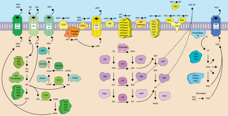
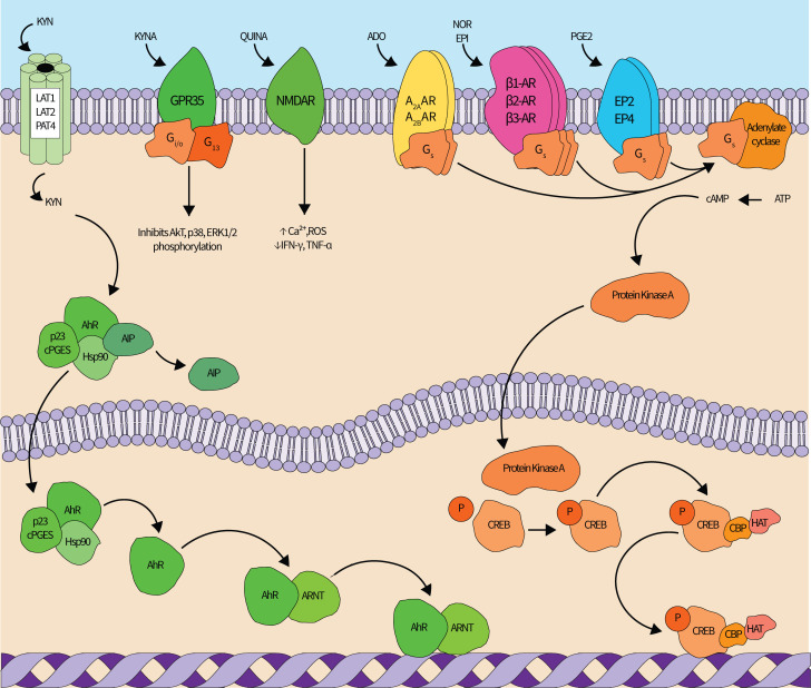
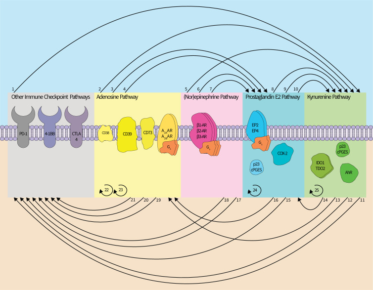
Tumors accumulate metabolites that deactivate infiltrating immune cells and polarize them toward anti-inflammatory phenotypes. We provide a comprehensive review of the complex networks orchestrated by several of the most potent immunosuppressive metabolites, highlighting the impact of adenosine, kynurenines, prostaglandin E2, and norepinephrine and epinephrine, while discussing completed and ongoing clinical efforts to curtail their impact. Retrospective analyses of clinical data have elucidated that their activity is negatively associated with prognosis in diverse cancer indications, though there is a current paucity of approved therapies that disrupt their synthesis or downstream signaling axes. We hypothesize that prior lukewarm results may be attributed to redundancies in each metabolites' synthesis or signaling pathway and highlight routes for how therapeutic development and patient stratification might proceed in the future.

Keywords: adenosine; immunotherapy; indoleamine-pyrrole 2,3-dioxygenase; metabolic networks and pathways; tumor escape.


Conflict of interest statement

Competing interests: JB has IP related to PEG-KYNase enzymes and has received consulting income from Ikena Oncology. DM has IP interests in the therapeutic use of IDO inhibitors; and has received consulting income and research support from NewLink Genetics/Lumos Pharma. MRJ is supported by a National Science Foundation Graduate Research Fellowship. DM receives funding from the NIH (R01CA103320 and R01CA211229). JB receives funding from the Emory University Winship Cancer Center and the Arnold and Mabel Beckman Foundation.

Figures

Figure 1
Figure 1
The redundant synthesis networks of immunosuppressive metabolites. Kynurenine pathway (left—green). After being imported into a cell by amino acid transporters, tryptophan is oxidized by one of three enzymes—IDO1, IDO2, or TDO2—to n-formyl-L-kynurenine, which is then converted to kynurenine by formamidases. Kynurenine may exit the cell through the LAT1, which simultaneously imports tryptophan, or continue down the kynurenine pathway until converted into xanthurenic acid (XANA) (major route) or nicotinamide adenine dinucleotide (NAD+) (minor route). Four different kynurenine aminotransferase (KAT) enzymes can transaminate kynurenine into kynurenic acid. Alternatively, kynurenine can be converted into 3-hydroxy-kynurenine by kynurenine 3-hydroxylase (K-3-H), or into anthranilic acid by kynureninase. 3-hydroxy-kynurenine may be converted to XANA (by KATs) or 3-hydroxyanthranilic acid (3-OHA) by kynureninase. 3-OHA may also be produced from anthranilic acid by 3-hydroxyanthranilic acid 3,4-hydroxylase (3 H-3,4-H). 3-OHA is converted into quinolinic acid by 3-hydroxyanthranilic acid 3,4-dioxygenase (3 H-3,4-D), and quinolinic acid phosphoribosyl transferase mediates the conversion of quinolinic acid to NAD+. XANA and kynurenic acid can be transported out of the cell by the OAT1 and OAT3, while connexin 43 allows NAD+ transport. Adenosine synthesis pathways (top center—yellow): adenosine synthesis occurs extracellularly in the tumor microenvironment and can use either ATP or NAD+ as a pathway substrate. ATP (or ADP to a lesser extent) can be dephosphorylated by CD39 or 6 other NTPDases into AMP. NAD+ can be converted to AMP directly by CD203a or through an ADP ribose (ADPR) intermediate by CD38 (followed by conversion to AMP by CD203a). AMP can also be generated from cAMP. cAMP is synthesized from protein Gs-activated adenylate cyclase from ATP. Intracellular cAMP can be excreted through multidrug resistance proteins 4, 5, or 8 into the extracellular space where a family of ecto-phosphodiesterases convert cAMP into AMP. AMP is finally dephosphorylated to adenosine by six possible enzymes, most prominently CD73. Adenosine can be deaminated to produce inosine by adenosine deaminase 1 (ADA) (membrane-attached via CD26) or 2. AMP can also be deaminated into IMP by AMP deaminase, and IMP can be dephosphorylated by CD73 to generate inosine (not shown). (Nor)epinephrine synthesis pathways (bottom center—magenta): schematic adapted from Molinoff and Axelrod. Epinephrine synthesis begins with tyrosine (TYR) and typically proceeds through L-dopa, dopamine (DOP), and norepinephrine (NOR) intermediates, though a parallel biosynthetic pathway exits. (Nor)epinephrine are exported through an undetermined mechanism. Prostaglandin E2 synthesis pathways (right—Blue): phospholipase A2s cleave AA from the inner leaflet of the cell membrane. Free AA is converted to PGH2 by the cyclooxygenase enzymes (COX-1, COX-2, and in the brain, COX-3). PGH2 is then converted to PGE2 by mPGES-1, mPGES-2, or cPGES (or p23), though PGH2 is also a precursor for several other prostaglandin derivatives (indicated by multiple reaction arrows stemming from PGH2 into the cytosol). PGE2 can also be generated when 8-iso-PGE2 undergoes epimerization. PGE2 is exported by MRP 1, MRP 2, or MRP 4. All proteins are denoted by text within shapes while metabolites are free-floating text. Straight lines indicate an enzyme catalyzed reaction, curved lines indicate transport or are drawn for clarity, and curved dotted lines indicate an undetermined transport mechanism. Stacked proteins indicate the existence of more than one enzyme capable of performing the indicated metabolic reaction. Up to 19 phospholipase enzymes exist in mammals, but only 1 is shown for clarity. 3-OHK, 3-hydroxykynurenine; AA, arachidonic acid; AAD, aromatic acid decarboxylase; ADO, adenosine; ANTHA, anthranilic acid; cAMP, cyclic AMP; CD, cluster of differentiation; CFE, catecholamine-forming enzyme; cPGES, cytosolic prostaglandin E synthase; DBH, dopamine-beta-hydroxylase; EPI, epinephrine; GCAP, germ cell alkaline phosphatase; IDO, indoleamine 2,3-dioxygenase; IAP, intestinal alkaline phosphatase; IMP, inosine monophosphate; INO, inosine; KYNA, kynurenic acid; KYN, kynurenine; KYNase, kynureninase; LAT, L-type/large neutral amino acid transporter; mPGES, microsomal PGE synthase; MRP, multidrug resistance-associated protein; NFL KYN, N-formyl-L-KYN; NM EPI, N-methyl epinephrine; NMT, non-specific methyltransferase; NTPDase, ectonucleoside triphosphate diphosphohydrolase; OAT, organic ion transporter; OCT, octopamine; PDE, phosphodiesterase; PGH2, prostaglandin H2; PLAP, placental alkaline phosphatase; PNMT, phenylethanolamine-N-methyltransferase; QAPT, quinolinic acid phosphoribosyl transferase; QUINA, quinolinic acid; SYN, synephrine; TDO, tryptophan 2,3-dioxygenase; TNAP, tissue-non-specific alkaline phosphatase; TRACP, tartrate-resistant acid phosphatase; TRP, tryptophan; TYRA, tyramine.
Figure 2
Figure 2
The redundant signaling networks of immunosuppressive metabolites. Kynurenine (KYN) pathway (left—green): KYN enters the cell via the LAT1, LAT2 or PAT4 transporters. In the cytoplasm, KYN binds the AhR-cPGES-Hsp90-AIP complex. AIP dissociates from the complex after KYN binds, and the KYN-AhR-cPGES-Hsp90 complex translocates into the nucleus. within the nucleus, cPGES and Hsp90 dissociate, and AhR forms a heterodimer with Arnt that modulates gene expression in a widespread manner. Kynurenic acid (KYNA) also agonizes the extracellular GPR35 receptor, which inhibits Akt, p38, and ERK1/2 phosphorylation, and quinolinic acid (QUINA) agonizes the NMDA receptors to increase intracellular Ca2+ and ROS and decrease IFN-γ and TNF-α production. Adenosine (ADO) (top center left—yellow): ADO can agonize four different G-protein coupled receptors (GPR35), of which A2AAR and A2BAR stimulate cAMP generation and downstream immunosuppression mediated by the cAMP-PKA-CREB pathway, shown in orange. (Nor)epinephrine (NOR) (top center right—magenta): NOR and epinephrine (EPI) can agonize either α-adrenergic or β-adrenergic receptors (β-AR). Agonism of all three β-AR elicits immunosuppressive signaling cascades through cAMP-PKA-CREB. Prostaglandin E2 (PGE2) (top rightadrenergic—blue): similar to ADO, PGE2 can agonize four receptors, of which two (EP2 and EP4) elicit immunosuppressive signaling cascades through cAMP-PKA-CREB. cAMP-PKA-CREB signaling pathway: cAMP production is increased by ADO, NOR, and/or PGE2 signaling through Gs-mediated activation of adenylate cyclase. adenylate cyclase generates cAMP from ATP, and cAMP then activates PKA. A cAMP-PKA complex enters the nucleus where it phosphorylates the CREB transcription factor. phosphorylated CREB recruits HAT and CBP and then modulates target gene expression. All proteins are denoted by text within shapes while metabolites are free-floating text. Straight lines indicate an enzyme catalyzed reaction while curved lines indicate transport or are included for clarity. AXAR, adenosine receptor; AhR, aryl hydrocarbon receptor; AIP, aryl hydrocarbon receptor-interacting protein; ARNT, aryl hydrocarbon receptor nuclear translocator; Ca2+, calcium ion; cAMP, cyclic AMP; CBP, CREB binding protein; cPGES, cytosolic PGE synthase; CREB, cAMP-response element binding protein; EPX, PGE2 receptor; ERK, extracellular signal-regulated kinase; GX, protein G; HAT, histone acetyltransferase; Hsp, heat shock protein; IFN-γ, interferon gamma; LAT, L-type/large neutral amino acid transporter; NMDAR, N-Methyl-D-aspartate receptor; P, phosphate group; PAT, proton-assisted amino acid transporter; ROS, reactive oxidative species; TNF-α, tumor necrosis factor alpha.
Figure 3
Figure 3
Interactions within and between the immunosuppressive metabolite networks. The adenosine (ADO), kynurenine, (PGE2), (nor)epinephrine (NOR), and other immune checkpoint pathways interact and stimulate each other and may employ feedforward signaling/synthesis regimes. (1) Blockade of PD-1 or CTLA-4 appears to induce (IDO1) expression. (2) A2BAR signaling induces (IDO1) expression. (3, 4) CD39 and CD73 can promote synthesis of (PGE2), and A2AAR or A2BAR signaling can induce expression of COX-2. (5) β-AR signaling upregulates (IDO1) expression. (6, 7) β2-AR signaling has been linked to the upregulation of COX-2 and cPGES. (8, 9, 10) cPGES is a modulator of (AhR) activity, and COX-2 can influence expression of (TDO2) and (IDO1). (11, 12) (AhR) signaling can increase PD-1 expression and correlates with reduced efficacy of an αCTLA-4 antibody. (13) (AhR) signaling can promote expression of (CD39.) (14) (AhR) signaling can promote expression of COX-2. (15) EP2/4 signaling can increase PD-1 expression. (16) (PGE2) can induce expression of CD73.) (17, 18) β-adrenergic signaling correlates with diminished therapeutic efficacy of αPD-1 antibodies, and decreases the therapeutic efficacy of a 4-1BB blockade. (19, 20) (CD73) expression correlates with diminished therapeutic efficacy of αPD-1 antibodies and of an αCTLA-4 antibody. (21) CD38 is associated with tumorous resistance to anti-PD-1/PD-L1 antibody therapy. (22, 23) A2A/BAR signaling drives (CD73) expression, and A2AAR signaling increases (CD39) expression. (24) PGE2-EP2/4 signaling can upregulate COX-2 expression. (25) (AhR) signaling drives upregulation of (IDO1) expression. In total, direct links between each pathway have been described with the exception of a relationship between ADO and NOR. Colors denoting ADO, kynurenine, (PGE2), and NOR pathways follow (figures 1 and 2). For brevity, the distinct cell types in which these interactions were described are not included. Curved lines between pathways indicate an interpathway interaction whereas curved semicircles indicate intrapathway regulation. AXAR, adenosine receptor; AhR, aryl hydrocarbon receptor; β-AR, β-adrenergic receptor; CD, cluster of differentiation; COX, cyclooxygenase; cPGES, cytosolic prostaglandin E synthase; CTLA, cytotoxic T-lymphocyte-associated protein; EPI, epinephrine; PGE receptorIDO, indoleamine 2,3-dioxygenase; PD, programmed cell death receptor; TDO, tryptophan 2,3-dioxygenase.

Similar articles

Cited by

References

    1. Vinay DS, Ryan EP, Pawelec G, et al. . Immune evasion in cancer: mechanistic basis and therapeutic strategies. Semin Cancer Biol 2015;35 Suppl:S185–98. 10.1016/j.semcancer.2015.03.004 - DOI - PubMed
    1. Vaddepally RK, Kharel P, Pandey R, et al. . Review of indications of FDA-approved immune checkpoint inhibitors per NCCN guidelines with the level of evidence. Cancers 2020;12. 10.3390/cancers12030738. [Epub ahead of print: 20 03 2020]. - DOI - PMC - PubMed
    1. Soda K. The mechanisms by which polyamines accelerate tumor spread. J Exp Clin Cancer Res 2011;30:95. 10.1186/1756-9966-30-95 - DOI - PMC - PubMed
    1. PeÑarando J, Aranda E, RodrÍguez-Ariza A. Immunomodulatory roles of nitric oxide in cancer: tumor microenvironment says "NO" to antitumor immune response. Transl Res 2019;210:99–108. 10.1016/j.trsl.2019.03.003 - DOI - PubMed
    1. Romero-Garcia S, Moreno-Altamirano MMB, Prado-Garcia H, et al. . Lactate contribution to the tumor microenvironment: mechanisms, effects on immune cells and therapeutic relevance. Front Immunol 2016;7:52. 10.3389/fimmu.2016.00052 - DOI - PMC - PubMed

Publication types