Comparative Study
. 2021 Oct 19;8(6):e1094.
doi: 10.1212/NXI.0000000000001094. Print 2021 Nov.

Evaluation of Age-Dependent Immune Signatures in Patients With Multiple Sclerosis

Affiliations
Comparative Study

Evaluation of Age-Dependent Immune Signatures in Patients With Multiple Sclerosis

Melanie Eschborn et al. Neurol Neuroimmunol Neuroinflamm. .

Abstract

Background and objectives: In MS, an age-related decline in disease activity and a decreased efficacy of disease-modifying treatment have been linked to immunosenescence, a state of cellular dysfunction associated with chronic inflammation.

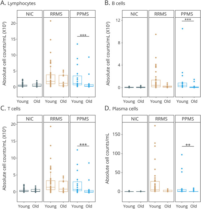
Methods: To evaluate age-related immunologic alterations in MS, we compared immune signatures in peripheral blood (PB) and CSF by flow cytometry in patients with relapsing-remitting (RR) (PB n = 38; CSF n = 51) and primary progressive (PP) MS (PB n = 40; CSF n = 36) and respective controls (PB n = 40; CSF n = 85).

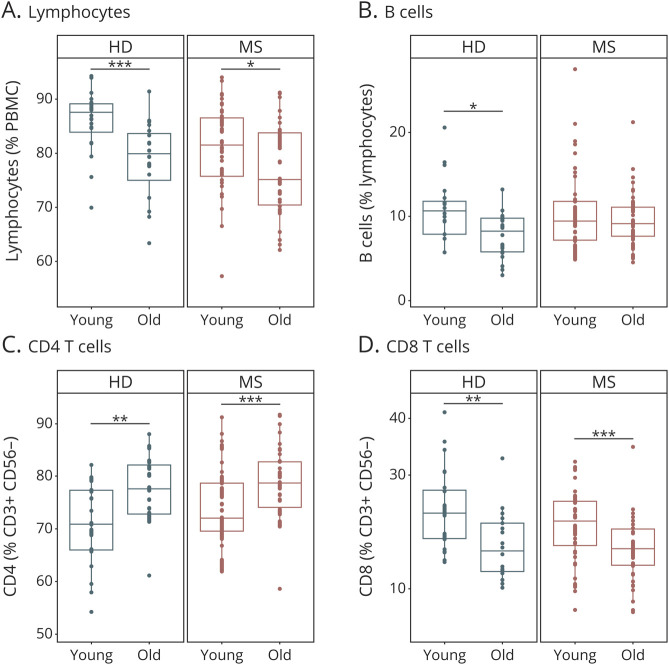
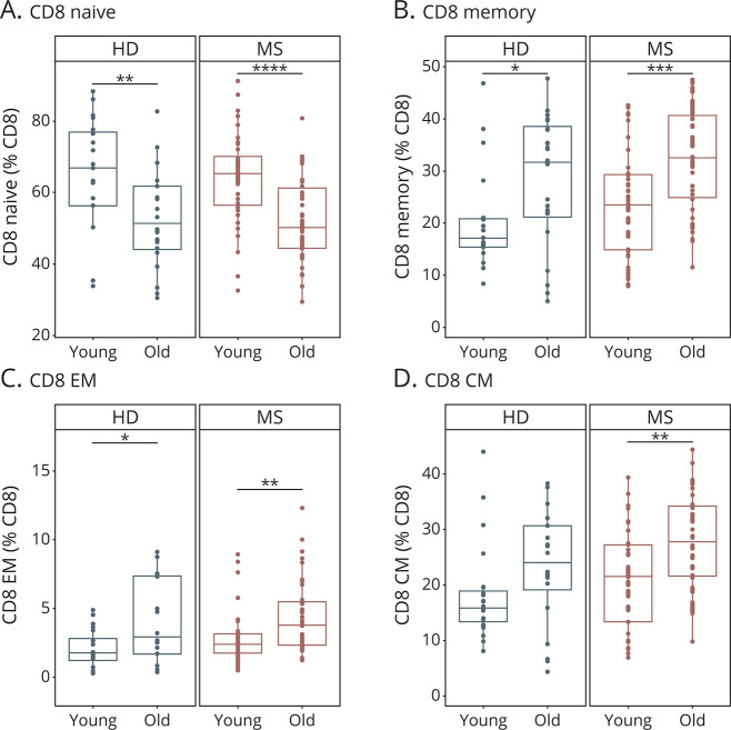
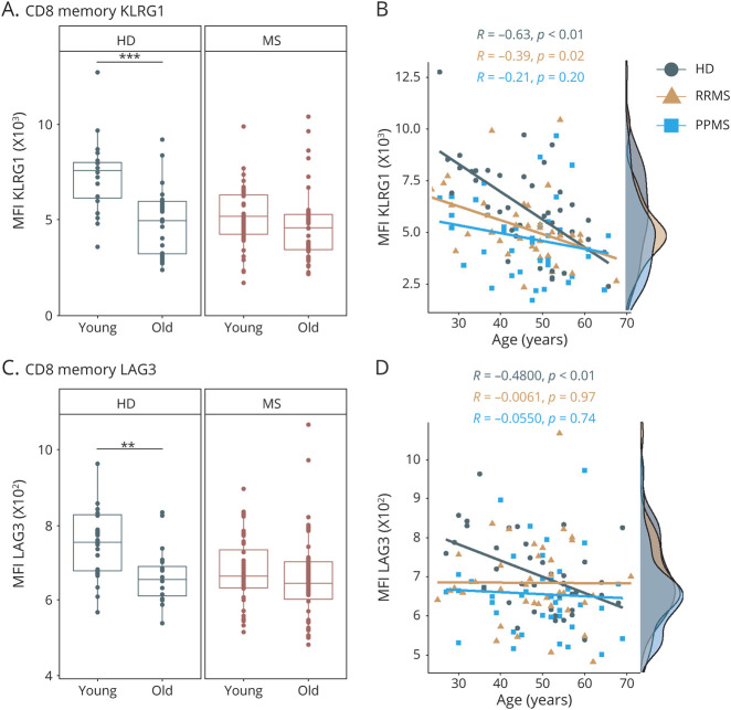
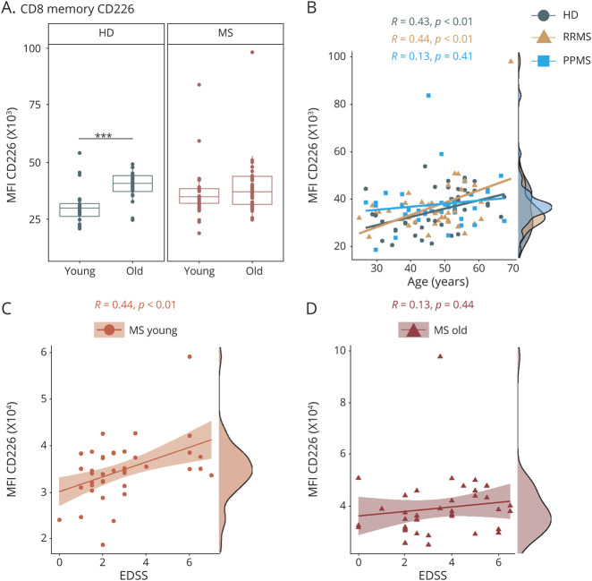
Results: Analysis revealed significant age-related changes in blood immune cell composition, especially in the CD8 T-cell compartment of healthy donors (HDs) and patients with MS. However, HDs displayed a strong age-dependent decline in the expression of the immunoregulatory molecules KLRG1, LAG3, and CTLA-4 on memory CD8 T cells, whereas this age-dependent reduction was completely abrogated in patients with MS. An age-dependent increase in the expression of the costimulatory molecule CD226 on memory CD8 T cells was absent in patients with MS. CD226 expression correlated with disability in younger (≤50 years) patients with MS. CSF analysis revealed a significant age-dependent decline in various immune cell populations in PPMS but not RRMS, suggesting a differential effect of aging on the intrathecal compartment in PPMS.

Discussion: Our data illustrate that aging in MS is associated with a dysbalance between costimulatory and immunoregulatory signals provided by CD8 T cells favoring a proinflammatory phenotype and, more importantly, a pattern of premature immune aging in the CD8 T-cell compartment of young patients with MS with potential implications for disease severity.


Figures

Figure 1
Figure 1. Basic Immune Cell Subset Composition in Young and Old Patients With MS and HDs
Immune cell subset composition in the peripheral blood of young (≤50 years) and old (>50 years) patients with multiple sclerosis (MS) (young: n = 40, old: n = 38) and HDs (young: n = 20, old: n = 20). Demographic data of study subjects are depicted in eTable 1 (A–D, links.lww.com/NXI/A620). Frequencies of lymphocytes (A), B cells (B), CD4 (C), and CD8 (D) T cells. Data are displayed as boxplots of the median and the 25th and 75th percentile ± interquartile range. Statistical analysis was conducted by the 2-tailed Mann-Whitney test. Differences were considered statistically significant with the following p values: *p < 0.05, **p < 0.01, and ***p < 0.001. HD = healthy donor.
Figure 2
Figure 2. Age-Related Changes in the CD8 T-Cell Compartment in Patients With MS and HDs
Composition of the CD8 T-cell compartment in the peripheral blood of young (≤50 years) and old (>50 years) patients with MS (young: n = 40, old: n = 38) and HDs (young: n = 20, old: n = 20). Demographic data of study subjects are depicted in eTable 1 (A–D, links.lww.com/NXI/A620). Proportions of naive (A), memory (B), effector memory (C), and central memory (D) CD8 T cells. Data are displayed as boxplots of the median and the 25th and 75th percentile ± interquartile range. Statistical analysis was conducted by the 2-tailed Mann-Whitney test. Differences were considered statistically significant with the following P values: *p < 0.05, **p < 0.01, ***p < 0.001, and ****p < 0.0001. CM = central memory; EM = effector memory; HD = healthy donor.
Figure 3
Figure 3. Immunoregulatory Markers on T Cells in Young and Old Patients With MS and HDs
Flow cytometric analysis of frozen peripheral blood mononuclear cells from young (≤50 years) and old (>50 years) patients with MS (MS: young: n = 40, old: n = 38; RRMS: young: n = 20, old: n = 18; PPMS: young: n = 20, old: n = 20) and HDs (young: n = 20, old: n = 20). Demographic data of study subjects are depicted in eTable 1, links.lww.com/NXI/A620. (A) MFI of KLRG1 on memory CD8 T cells of patients with MS and HDs. (B) Correlation analysis of MFI of KLRG1 on memory CD8 T cells with age of HDs (n = 40), patients with RRMS (n = 38), and patients with PPMS (n = 40). (C) MFI of LAG3 on memory CD8 T cells of patients with MS and HDs. (D) Correlation analysis of MFI of LAG3 on memory CD8 T cells with age of HDs (n = 40), patients with RRMS (n = 38), and patients with PPMS (n = 40). Data are displayed as boxplots of the median and the 25th and 75th percentile ± interquartile range. Statistical analysis was conducted by the 2-tailed Mann-Whitney test. For correlation analysis, the Pearson product-moment correlation coefficients (Pearson R) were computed. Differences were considered statistically significant with the following p values: **p < 0.01 and ***p < 0.001. HD = healthy donor; KLRG1 = killer cell lectin-like receptor subfamily G member 1; LAG3 = lymphocyte-activation gene 3; MFI = mean fluorescence intensity; MS = multiple sclerosis; PPMS = primary progressive MS; RRMS = relapsing-remitting MS.
Figure 4
Figure 4. Age-Related Expression of the Costimulatory Molecule CD226 on T Cells in Patients With MS and HDs
Flow cytometric analysis of frozen peripheral blood mononuclear cells from young (≤50 years) and old (>50 years) patients with MS (MS: young: n = 40, old: n = 38; RRMS: young: n = 20, old: n = 18; PPMS: young: n = 20, old: n = 20) and HDs (young: n = 20, old: n = 20). Demographic data of study subjects are depicted in eTable 1, links.lww.com/NXI/A620. (A) MFI of CD226 (DNAM-1) on memory CD8 T cells. (B) Correlation analysis of MFI of CD226 on memory CD8 T cells with age of HDs (n = 40), patients with RRMS (n = 38), and patients with PPMS (n = 40). (C, D) Correlation analysis of CD226 expression on memory CD8 T cells with the EDSS score of young (C) and old (D) patients with MS. Data are displayed as boxplots of the median and the 25th and 75th percentile ± interquartile range. Statistical analysis was conducted by the 2-tailed Mann-Whitney test. For correlation analysis, the Pearson product-moment correlation coefficients (Pearson R) were computed. Differences were considered statistically significant with the following p values: **p < 0.01 and ***p < 0.001. EDSS = Expanded Disability Status Scale; HD = healthy donor; MS = multiple sclerosis; MFI = mean fluorescence intensity; PPMS = primary progressive MS; RRMS = relapsing-remitting MS.
Figure 5
Figure 5. Age-Dependent Immune Cell Subset Composition in the CSF of Patients With MS and Noninflammatory Controls
Immune cell subset composition in the in the CSF of young (≤50 years) and old (>50 years) NICs (young n = 60; old: n = 25) and patients with RRMS (young: n = 41; old: n = 10) and PPMS (young: n = 15; old: n = 21). Demographic data of study subjects are depicted in eTable 2, links.lww.com/NXI/A621. (A–D) Absolute numbers of lymphocytes (A), B cells (B), T cells (C), and plasma cells (D). Data are displayed as boxplots of the median and the 25th and 75th percentile ± interquartile range. Statistical analysis was conducted by the 2-tailed Mann-Whitney test. Differences were considered statistically significant with the following p-values: **p < 0.01 and ***p < 0.001. NIC = noninflammatory control; PPMS = primary progressive MS; RRMS = relapsing-remitting MS.

References

    1. Pawelec G. Age and immunity: What is “immunosenescence”? Exp Gerontol. 2018;105:4-9. - PubMed
    1. Ray D, Yung R. Immune senescence, epigenetics and autoimmunity. Clin Immunol. 2018;196:59-63. - PMC - PubMed
    1. Vaughn CB, Jakimovski D, Kavak KS, et al. Epidemiology and treatment of multiple sclerosis in elderly populations. Nat Rev Neurol. 2019;15(6):329-342. - PubMed
    1. Weideman AM, Tapia-Maltos MA, Johnson K, Greenwood M, Bielekova B. Meta-analysis of the age-dependent efficacy of multiple sclerosis treatments. Front Neurol. 2017;8:577-612. - PMC - PubMed
    1. Sanai SA, Saini V, Benedict RH, et al. Aging and multiple sclerosis. Mult Scler. 2016;22(6):717-725. - PubMed

Publication types

MeSH terms