. 2021 Oct 19;12(11):966.
doi: 10.1038/s41419-021-04263-w.

Loss of Numb promotes hepatic progenitor expansion and intrahepatic cholangiocarcinoma by enhancing Notch signaling

Affiliations

Loss of Numb promotes hepatic progenitor expansion and intrahepatic cholangiocarcinoma by enhancing Notch signaling

Yuke Shu et al. Cell Death Dis. .

Abstract

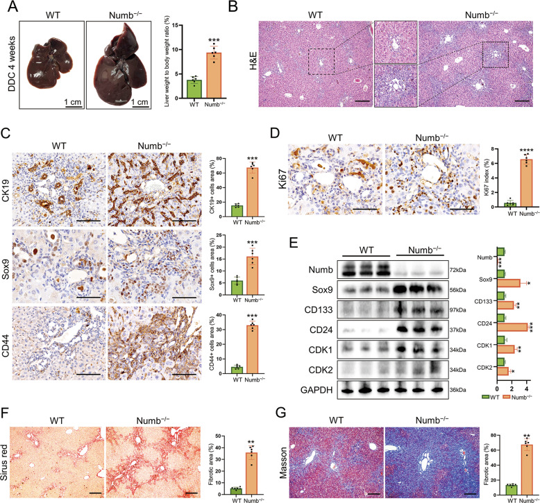
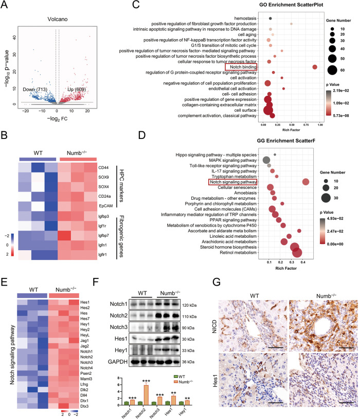
Numb, a stem cell fate determinant, acts as a tumor suppressor and is closely related to a wide variety of malignancies. Intrahepatic cholangiocarcinoma (iCCA) originates from hepatic progenitors (HPCs); however, the role of Numb in HPC malignant transformation and iCCA development is still unclear. A retrospective cohort study indicated that Numb was frequently decreased in tumor tissues and suggests poor prognosis in iCCA patients. Consistently, in a chemically induced iCCA mouse model, Numb was downregulated in tumor cells compared to normal cholangiocytes. In diet-induced chronic liver injury mouse models, Numb ablation significantly promoted histological impairment, HPC expansion, and tumorigenesis. Similarly, Numb silencing in cultured iCCA cells enhanced cell spheroid growth, invasion, metastasis, and the expression of stem cell markers. Mechanistically, Numb was found to bind to the Notch intracellular domain (NICD), and Numb ablation promoted Notch signaling; this effect was reversed when Notch signaling was blocked by γ-secretase inhibitor treatment. Our results suggested that loss of Numb plays an important role in promoting HPC expansion, HPC malignant transformation, and, ultimately, iCCA development in chronically injured livers. Therapies targeting suppressed Numb are promising for the treatment of iCCA.


Conflict of interest statement

The authors declare no competing interests.

Figures

Fig. 1
Fig. 1. Numb is downregulated in human iCCA and mouse iCCA.
A Immunohistochemistry staining of Numb expression in iCCA specimens and paired normal tissues. CK19 staining confirmed the cholangiocyte origins of the tumors. Scale bar, 50 μm. B Western blotting showing the different Numb protein levels between two pairs of iCCA tissues and adjacent nonmalignant tissues. The corresponding statistical analysis is also presented. C, D Correlation of Numb expression with the recurrence-free survival (RFS) time and the overall survival (OS) time of iCCA patients, respectively. E Immunofluorescence staining and F Western blotting showing that Numb was downregulated in mouse iCCA tissues (TAA treated for 6 months) with the corresponding statistical analysis. Scale bars, 50 μm. Data represent the mean ± SD of at least three independent experiments; *P < 0.05, **P < 0.01, and ***P < 0.001.
Fig. 2
Fig. 2. Numb ablation promotes HPC expansion and liver fibrosis.
A Gross views and the liver weight/body weight ratios of WT and Numb−/− livers after 4 weeks of DDC exposure. B Histological alterations were assessed with H&E staining. Scale bar, 200 μm. C Expression of HPC markers (CK19, Sox9, and CD44) and corresponding statistical results. Scale bars, 100 μm (CK19 and CD44) and 50 μm (Sox9). D Ki67 immunohistochemistry staining showing the active proliferation of HPCs in Numb−/− livers. Scale bar, 50 μm. E Western blotting showing elevated progenitor markers (CD133, Sox9, and CD24) and critical cell cycle proteins (CDK1 and CDK2), and the relative expression of these proteins was also semiquantified and analyzed. F, G Masson trichrome and Sirius red staining were performed to measure liver fibrosis. Scale bar, 200 μm. All data represent the mean ± SD of at least three independent experiments; *P < 0.05, **P < 0.01, and ***P < 0.001.
Fig. 3
Fig. 3. Numb ablation promotes HPC expansion, periportal fibrosis, and iCCA.
A Gross views of WT and Numb−/− livers after 4 months of TAA exposure and the tumorigenesis ratio of WT and Numb−/− mice at specific times. The black arrowhead indicates the tumor nodule in the Numb−/− liver. B CK19 and HNF4α immunohistochemistry staining showing that the tumor was cholangiocarcinoma. Scale bars, 50 μm. C Masson trichrome and Sirius red staining were performed to measure liver fibrosis. Scale bar, 200 μm. D Histological alterations were assessed with H&E staining. Scale bar, 200 μm. E, F Immunohistochemistry staining of Sox9 and Ki67 in WT and Numb−/− livers, respectively. Scale bars, 25 μm. The corresponding statistical results are also shown. G Western blotting showing the elevated expression of progenitor markers (CD133, Sox9, and CD24) and critical cell cycle-related proteins (CDK1 and CDK2), and the relative expression of these proteins was also semiquantified and analyzed. All data represent the mean ± SD of at least three independent experiments; *P < 0.05, **P < 0.01, and ***P < 0.001.
Fig. 4
Fig. 4. Numb knockdown promotes proliferation, metastasis, and stemness in iCCA cells.
The colony formation assay (A), migration assay (B), and wound-healing assay (C) were performed in HuCCT1 and RBE cell lines. D Progenitor markers (CD133, Sox9, and CD24) and critical cell cycle proteins (CDK1 and CDK2) were upregulated after Numb silencing in HuCCT1 and RBE cells. E Sphere formation assays comparing the stemness of HuCCT1 and RBE cells after Numb silencing. Scale bar, 100 μm. F Immunofluorescence staining showing the upregulation of progenitor markers CD133 and Sox9 after Numb silencing. Scale bars, 50 μm (CD133) and 100 μm (Sox9). All data represent the mean ± SD of at least three independent experiments; *P < 0.05, **P < 0.01, and ***P < 0.001.
Fig. 5
Fig. 5. Numb deficiency promotion of iCCA is dependent on Notch signaling.
A Volcano plot displaying the differentially expressed genes between WT and Numb−/− mouse nontumorous liver tissues. B Expression of HPC markers and profibrogenic genes. C, D GO analysis and KEGG analysis showing that the differentially expressed genes were enriched in the Notch signaling pathway. E Alterations in the Notch signaling pathways between WT livers and Numb−/− livers after TAA treatment. F Western blotting analysis of Notch-associated proteins and the key downstream proteins of Notch, NICD, and Hes1. G NICD and Hes1 immunohistochemistry staining in WT and Numb−/− mouse livers. Scale bar, 50 μm. All data represent the mean ± SD of at least three independent experiments; *P < 0.05, **P < 0.01, and ***P < 0.001.
Fig. 6
Fig. 6. Numb regulates Notch signaling by binding to NICD in vitro.
A Proliferation, metastasis, and stemness were measured by colony formation, migration, wound-healing, and sphere formation assays after DAPT treatment at a dose of 150 μM (the control group was treated with the same volume of DMSO without DAPT) in HuCCT1 cells. Scale bar, 200 μm. B Immunofluorescence staining of HPC markers (CD133 and Sox9) in HuCCT1 cells. Scale bars, 50 μm (CD133) and 100 μm (Sox9). C Immunofluorescence staining of the key proteins of Notch signaling, NICD, and Hes1, in HuCCT1 cells. Scale bar, 50 μm. D Western blotting displaying the upregulation of HPC markers and the downstream factors of Notch after Numb silencing was reversed after DAPT treatment in HuCCT1 cells. E Coimmunoprecipitation (CoIP) showing the direct binding of Numb and NICD. All data represent the mean ± SD of at least three independent experiments; *P < 0.05, **P < 0.01, and ***P < 0.001.
Fig. 7
Fig. 7. Negative correlation between Numb and NICD expression in human iCCA.
A Immunohistochemistry staining of Numb, NICD, Hes1, and Ki67 in NumbHigh iCCA samples and NumbLow iCCA samples. Scale bar, 100 μm. B Diagram of the hypothetical roles of Numb in HPC expansion, liver fibrosis, and iCCA development by regulating the Notch signaling pathway. All data represent the mean ± SD of at least three independent experiments; *P < 0.05, **P < 0.01, and ***P < 0.001.

References

    1. Sung H, Ferlay J, Siegel RL, Laversanne M, Soerjomataram I, Jemal A, et al. Global cancer statistics 2020: GLOBOCAN estimates of incidence and mortality worldwide for 36 cancers in 185 countries. CA Cancer J Clin. 2021;71:209–49. doi: 10.3322/caac.21660. - DOI - PubMed
    1. Bertuccio P, Bosetti C, Levi F, Decarli A, Negri E, La, et al. A comparison of trends in mortality from primary liver cancer and intrahepatic cholangiocarcinoma in Europe. Ann Oncol. 2013;24:1667–74. doi: 10.1093/annonc/mds652. - DOI - PubMed
    1. Razumilava N, Gores GJ. Cholangiocarcinoma. Lancet. 2014;383:2168–79. doi: 10.1016/S0140-6736(13)61903-0. - DOI - PMC - PubMed
    1. Rizvi S, Khan SA, Hallemeier CL, Kelley RK, Gores GJ. Cholangiocarcinoma - evolving concepts and therapeutic strategies. Nat Rev Clin Oncol. 2018;15:95–111. doi: 10.1038/nrclinonc.2017.157. - DOI - PMC - PubMed
    1. Fujita T. Liver transplantation for intrahepatic cholangiocarcinoma. Lancet. 2014;384:1182. doi: 10.1016/S0140-6736(14)61716-5. - DOI - PubMed

Publication types

MeSH terms