. 2021 Oct 19;12(1):6086.
doi: 10.1038/s41467-021-26366-w.

Structural basis of soluble membrane attack complex packaging for clearance

Affiliations

Structural basis of soluble membrane attack complex packaging for clearance

Anaïs Menny et al. Nat Commun. .

Abstract

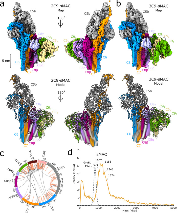
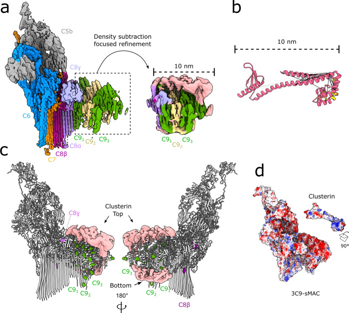
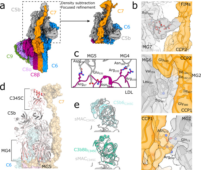
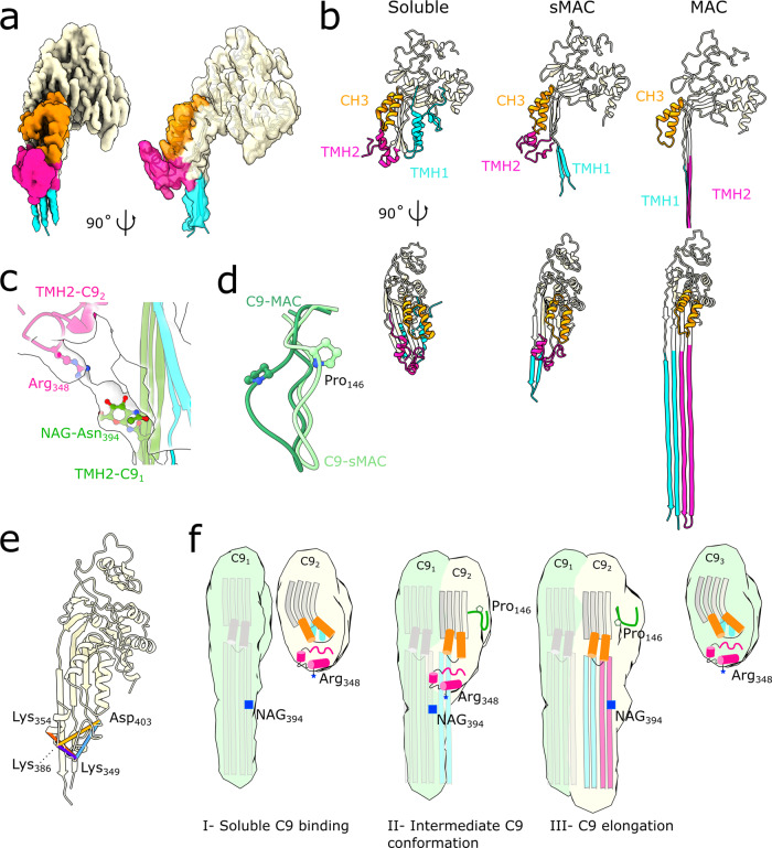
Unregulated complement activation causes inflammatory and immunological pathologies with consequences for human disease. To prevent bystander damage during an immune response, extracellular chaperones (clusterin and vitronectin) capture and clear soluble precursors to the membrane attack complex (sMAC). However, how these chaperones block further polymerization of MAC and prevent the complex from binding target membranes remains unclear. Here, we address that question by combining cryo electron microscopy (cryoEM) and cross-linking mass spectrometry (XL-MS) to solve the structure of sMAC. Together our data reveal how clusterin recognizes and inhibits polymerizing complement proteins by binding a negatively charged surface of sMAC. Furthermore, we show that the pore-forming C9 protein is trapped in an intermediate conformation whereby only one of its two transmembrane β-hairpins has unfurled. This structure provides molecular details for immune pore formation and helps explain a complement control mechanism that has potential implications for how cell clearance pathways mediate immune homeostasis.


Conflict of interest statement

The authors declare no competing interests.

Figures

Fig. 1
Fig. 1. sMAC is a complement activation macromolecule with a heterogeneous composition.
a CryoEM reconstruction (top) and atomic model (bottom) of sMAC that consists of a core complement complex (C5b, C6, C7, and C8α/C8β/C8γ) together with two C9 molecules (C91, C92). b CryoEM reconstruction (top) and atomic model (bottom) of sMAC with the same core complement complex and three molecules of C9 (C91, C92, C93). CryoEM density maps in a and b are colored according to protein composition. Glycans included in the atomic models are shown as sticks in the ribbon diagrams. c sMAC circos plot of identified 4-(4,6-Dimethoxy-1,3,5-triazin-2-yl)-4-methylmorpholinium chloride (DMTMM) cross-links within and across complement components (C5, C6, C7, C8, and C9). The complement components are cross-linked to the chaperones vitronectin (VTNC) and clusterin (CLUS). Intra-links are shown as orange lines and inter-links are shown as gray lines. Source data are provided as a Source Data file. d Mass photometry of sMAC reveals a heterogeneous mixture of complexes (orange line). Masses of the most abundant complexes are indicated. Gaussian distribution of GroEL from the protein standard is shown as a reference for peak width of a homogeneous complex (gray dotted line).
Fig. 2
Fig. 2. Clusterin bridges complement proteins in sMAC through electrostatic interactions.
a CryoEM reconstruction of 3C9-sMAC highlighting the region used in subsequent focused refinements (black-dotted lines). Inset shows the map after density subtraction of the core complement complex and refinement of the C9 oligomer. Density is colored according to protein composition, with regions of the map not accounted for by complement proteins in pink. b Structural homology model for the clusterin core (pink ribbons) with intra-molecular clusterin cross-links derived from XL-MS mapped (black lines). Over-length cross-links are shown as gray lines and known disulfide bonds within clusterin are shown in yellow. Source data are provided as a Source Data file. c Intermolecular cross-links between clusterin and C8/C9 plotted on the 3C9-sMAC model (white ribbons). Complement protein residues involved in cross-links are shown as spheres colored according to protein composition. Density not corresponding to complement proteins in the focused refined map (pink surface) is overlaid for reference. d Coulombic electrostatic potential ranging from −10 (red) to 10 (blue) kcal/(mol·e) calculated from the models for complement proteins in 3C9-sMAC (bottom surface) and the model for clusterin shown in 2b (top surface).
Fig. 3
Fig. 3. C7 connects conformational changes of the C345C domain with the MG scaffold of C5b.
a CryoEM reconstruction of 2C9-sMAC highlighting the region used in subsequent focused refinements (black-dotted lines). Inset corresponds to the map resulting from density subtraction of the MACPF-arc and focused refinement on C5b. Density is colored by protein composition. b Panels show density from the focused refined map (colored by protein composition) overlaid with the sMAC atomic model (ribbons) at three interaction interfaces between C7 (orange) and C5b (gray). Glycan extending from C5b:Asn893 stabilizes a linker between the C-terminal CCP and first FIM domain of C7 (top). C5b:Tryp581 locks into a hydrophobic hinge between the CCP1 and CCP2 domains of C7 (middle). Ionic interactions between the first CCP of C7 and the MG1 domain of C5b (bottom). Side-chains of interface residues shown as sticks. c Interface between the C8β LDL domain (purple) and C5b MG scaffold (MG4 and MG5 in grey). Side-chains of interface residues are shown as sticks. d C5b within sMAC (ribbons) colored by RMSD with superposed C5b from the C5b6 crystal structure (PDB ID: 4A5W). Red indicates residues with maximal differences. C345C, MG4 and MG5 domains of C5b are highlighted. C6 (blue), C8 (pink) and C7 (orange) are shown as semi-transparent surfaces for reference. e Superposition of C5b within sMAC (grey) with corresponding residues in the soluble C5b6 complex (cyan) (PDB ID: 4A5W) showing movement of the C345C domain (top panel). Superposition of C5b from sMAC (grey) with the structural homolog C3b (green) from the C3b:Bb:Properdin complex (PDB ID: 6RUR). In both panels, alignments were done on the full molecule and C345C domains were cropped for clarity.
Fig. 4
Fig. 4. sMAC traps an alternative conformation of C9.
a Density for the terminal C9 in the 2C9-sMAC reconstruction (EMD-12647). Density corresponding to the CH3 (orange), TMH2 (pink) and TMH1 (cyan) regions of the MACPF domain are highlighted. The remainder of C9 is beige. Model for the alternative C9 conformation is overlaid (ribbon). b Ribbon diagrams for three conformations of C9: soluble C9 from the murine crystal structure (PDB ID: 6CXO) left panel, terminal C9 in 2C9-sMAC, and transmembrane conformation of C9 in MAC (PDB ID: 6H03). CH3, TMH1, and TMH2 regions of the MACPF domain are colored as in a. For the MAC conformation, the full length of C9 TMH hairpins are shown in the bottom right panel. c Interaction between the penultimate C9 TMH2 β-hairpins (green) with the helical TMH2 of the terminal C9 (pink). Side-chains of interface residues (NAG-Asn394 and Arg348) are shown as sticks. TMH1 of the terminal C9 (cyan) is shown for context. Density for this region is shown as a transparent surface. d Superposition of the C9 MACPF from MAC (dark green) and the conformation in the terminal C9 (light green) shows differences in a loop that contains the disease affected residue Pro146 (P146S). e Cross-links between TMH2 residues (Lys354, Lys386, Lys349, Asp403) derived from XL-MS and mapped on the terminal C9 conformation of 2C9-sMAC (beige ribbons). Distance lengths: 9 Å (purple), 14 Å (red), 17 Å (blue), 20 Å (yellow) are shown. Source data are provided as a Source Data file. f Schematic showing structural timeline of MACPF pore formation. In the soluble conformation (C92, C93), the MACPF domain comprises a central kinked β-sheet (grey) and the pore-forming residues within TMH1 (cyan) and TMH2 (pink) are helical. Upon binding the leading edge of the oligomer (C91), the central β-sheet straightens and two other MACPF regions: CH3 (orange cylinders) and a proline loop (dark green) rotate as TMH1 (cyan) is released. The intermediate conformation is stabilized by the interaction between a basic residue on TMH2 (Arg348, blue star) with a glycan on the β-strands of the preceding monomer (NAG-Asn394, blue square). This interaction may play a role in positioning TMH2 before the hairpins are sequentially released to propagate the pore.

Similar articles

Cited by

References

    1. Tomlinson S, Taylor PW, Morgan BP, Luzio JP. Killing of gram-negative bacteria by complement. Fractionation of cell membranes after complement C5b-9 deposition on to the surface of Salmonella minnesota Re595. Biochem. J. 1989;263:505–511. doi: 10.1042/bj2630505. - DOI - PMC - PubMed
    1. Brodsky RA. Paroxysmal nocturnal hemoglobinuria. Blood. 2014;124:2804–2811. doi: 10.1182/blood-2014-02-522128. - DOI - PMC - PubMed
    1. Morgan BP, Harris CL. Complement, a target for therapy in inflammatory and degenerative diseases. Nat. Rev. Drug Discov. 2015;14:857–877. doi: 10.1038/nrd4657. - DOI - PMC - PubMed
    1. Farkas I, et al. CD59 blocks not only the insertion of C9 into MAC but inhibits ion channel formation by homologous C5b-8 as well as C5b-9. J. Physiol. 2002;539:537–545. doi: 10.1113/jphysiol.2001.013381. - DOI - PMC - PubMed
    1. Doorduijn DJ, et al. Bacterial killing by complement requires direct anchoring of membrane attack complex precursor C5b-7. PLoS Pathog. 2020;16:e1008606. doi: 10.1371/journal.ppat.1008606. - DOI - PMC - PubMed

Publication types

MeSH terms