Review
. 2021 Oct 20;9(1):44.
doi: 10.1038/s41413-021-00168-8.

Roles of mechanosensitive channel Piezo1/2 proteins in skeleton and other tissues

Affiliations
Review

Roles of mechanosensitive channel Piezo1/2 proteins in skeleton and other tissues

Lei Qin et al. Bone Res. .

Abstract

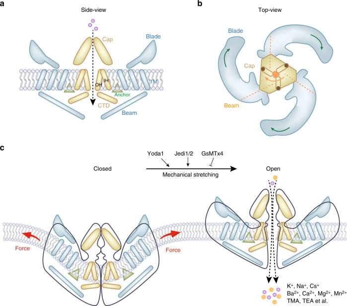
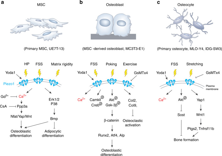
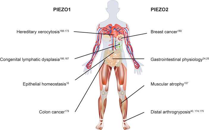
Mechanotransduction is a fundamental ability that allows living organisms to receive and respond to physical signals from both the external and internal environments. The mechanotransduction process requires a range of special proteins termed mechanotransducers to convert mechanical forces into biochemical signals in cells. The Piezo proteins are mechanically activated nonselective cation channels and the largest plasma membrane ion channels reported thus far. The regulation of two family members, Piezo1 and Piezo2, has been reported to have essential functions in mechanosensation and transduction in different organs and tissues. Recently, the predominant contributions of the Piezo family were reported to occur in the skeletal system, especially in bone development and mechano-stimulated bone homeostasis. Here we review current studies focused on the tissue-specific functions of Piezo1 and Piezo2 in various backgrounds with special highlights on their importance in regulating skeletal cell mechanotransduction. In this review, we emphasize the diverse functions of Piezo1 and Piezo2 and related signaling pathways in osteoblast lineage cells and chondrocytes. We also summarize our current understanding of Piezo channel structures and the key findings about PIEZO gene mutations in human diseases.


Conflict of interest statement

The authors declare no competing interests.

Figures

Fig. 1
Fig. 1
Mouse Piezo1 protein has a three-bladed, propeller-shaped homotrimeric architecture. a Side view of mouse Piezo1 channel. Piezo1 consists of a central ion-conducting pore modulus (yellow components) and the peripheral mechanotransduction modulus (blue component). The pore module contains the extracellular Cap structure, the transmembrane pore formed from three pairs of TMs, and the intracellular C-terminal domain (CTD). The peripheral mechanotransduction modulus includes a long beam-like structure, a peripheral blade, and a unique anchor domain. The anchor domain formed from a hairpin structure is connected to the CTD plane by the inner helix (IH) and outer helix (OH) pair, which maintains the integrity of the channel. The long beam structure supports and bridges the blade into the central pore module. b Top view of mouse Piezo1 channel. The large extracellular blade domains can curve the plasma membrane, and the three blades are assembled into functional trimers. c Mammalian Piezo1 proteins can be directly gated by membrane stretching, which is conserved throughout evolution. Yoda1 and Jedi1/2 are chemical activators of Piezo channels, and GsMTx4 is an antagonist of the Piezo1 channel. Piezo channels are nonselective cationic mechanosensitive channels that are permeable to alkali ions (K+, Na+, and Cs+), divalent cations (Ba2+, Ca2+, Mg2+, and Mn2+), and several organic cations (tetramethyl ammonium (TMA), tetraethyl ammonium (TEA)). Illustrations were modified from Wang et al. and Jiang et al.
Fig. 2
Fig. 2
Osteocyte Piezo1 deficiency leads to significant bone loss. a, b Micro-CT scanning of distal femurs from 3-month-old control (Piezo1fl/fl) and conditional knockout (cKO) mice with specific Piezo1 loss in osteocytes (Piezo1Dmp1). a’, b’ Cross-section of CT scan images at the red line in a, b for trabecular and cortical bone mass detection in Control and cKO mice. c, d Rhodamine-phalloidin staining of the F-actin cytoskeleton of cross-section samples of femurs from 3-month-old control and cKO mice. F-actin in green; DAPI in blue
Fig. 3
Fig. 3
Piezo1 signaling in osteoblast lineage cells. a The activation of Piezo1 channels in primary MSCs or UE7T-13 cell lines can be triggered by hydrostatic pressure (HP), fluid shear stress (FSS), matrix rigidity, and Yoda1, which further activate the Ppp3a-Nfat/Yap-Wnt pathway through Ca2+ signals and Bmp signals through the Erk1/2-p38 pathways. The activation of Piezo1 in MSCs leads to osteoblastic differentiation and inhibition of adipocytic differentiation. b Mechanical stimuli, such as FSS, poking, and exercise, induce Piezo1 activation in MSC-derived osteoblasts and MC3TC-E1 cell lines. Piezo1 activation stimulates intracellular signal responses, including Ca2+ flux, CamKII-Creb signaling, Akt-Gsk3β signaling, and collagen type 2 and 9 expression in osteoblasts. As a result, Piezo1 activation enhances osteoblastic differentiation but inhibits osteoclastic activation. c Piezo1 channels from primary osteocytes, MLO-Y4 or IDG-SW3 cell lines can be triggered by FSS and stretching stimuli. The activation of Piezo1 further induces Ca2+ flux, phosphorylation of Akt, and Yap1 activation in osteocytes. As a result, Piezo1 channels contribute to bone formation in these cells
Fig. 4
Fig. 4
Piezo1 signaling in chondrocytes. Under pathological mechanical loading, the expression of Piezo1 protein is increased, resulting in excessive Ca2+ influx. Ca2+ overload activates endoplasmic reticulum (ER) stress and upregulates the expression of caspase-12 (Casp-12), leading to the expression of caspase-3/7 (Casp-3/7), Bax, and Bcl2 and the release of cytochrome c (Cytc). Cytc and Apaf-1 upregulate the expression of caspase-9 (Casp-9), which activates Casp-3/7 to cleave Casp-3/7 (cCasp-3/7) and finally results in chondrocyte apoptosis. GsMTx4 and urocortin1 can exert chondroprotective effects by inhibiting Piezo1 and preventing Ca2+ overload
Fig. 5
Fig. 5
Piezo studies in mice. Piezo1 is widely expressed in multiple tissues with a preference for the endothelium. Studies focused on tissue-specific deletion of Piezo1 in experimental mice demonstrate the importance of Piezo1 in regulating lung development, angiogenesis, blood pressure control, bone development, lymphatic valve function, heart development and adipocyte differentiation and pancreas functions. Piezo2 is highly expressed in neurons. Current studies reveal the great contribution of Piezo2 to regulating mechanotransduction in central nervous system (CNS) neurons, dorsal root ganglia (DRG) neurons, and other sensory neurons
Fig. 6
Fig. 6
Piezo proteins in health and diseases in humans. In healthy humans, Piezo proteins play an important role in epithelial homeostasis and gastrointestinal physiology. Under pathological conditions, loss-of-function mutations in the human PIEZO1 gene are linked to autosomal-recessive congenital generalized lymphatic dysplasia of Fotiou (GLDF). Gain-of-function mutations in the human PIEZO1 gene cause hereditary xerocytosis (HX). Loss-of-function mutations in the human PIEZO2 gene result in an autosomal-recessive syndrome of muscular atrophy. Gain-of-function mutations in the human PIEZO2 gene lead to autosomal-dominant distal arthrogryposis (DA). In addition to hereditary human diseases, abnormal PIEZO protein expression is associated with colon cancer and breast cancer

Similar articles

Cited by

References

    1. Árnadóttir J, Chalfie M. Eukaryotic mechanosensitive channels. Annu. Rev. Biophys. 2010;39:111–137. - PubMed
    1. Duscher D, et al. Mechanotransduction and fibrosis. J. Biomech. 2014;47:1997–2005. - PMC - PubMed
    1. Romani P, Valcarcel-Jimenez L, Frezza C, Dupont S. Crosstalk between mechanotransduction and metabolism. Nat. Rev. Mol. Cell Biol. 2021;22:22–38. - PubMed
    1. Vollrath MA, Kwan KY, Corey DP. The micromachinery of mechanotransduction in hair cells. Annu. Rev. Neurosci. 2007;30:339–365. - PMC - PubMed
    1. Lyon Robert C, Zanella F, Omens Jeffrey H, Sheikh F. Mechanotransduction in cardiac hypertrophy and failure. Circ. Res. 2015;116:1462–1476. - PMC - PubMed