

Unusual Presentation of Lipofibromatosis-Like Neural Tumor in an Adult: A Case Report
- PMID: 34667475
- PMCID: PMC8473993
- DOI: 10.4103/sjmms.sjmms_63_21
Unusual Presentation of Lipofibromatosis-Like Neural Tumor in an Adult: A Case Report
Abstract
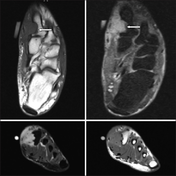
Lipofibromatosis-like neural tumor (LPF-NT) is a rare variant of lipofibromatosis. Standard lipofibromatosis tumors show a predilection for the distal extremities of male children or young adults and are typically painless, slow-growing, subcutaneous or deep soft tissue masses. We present a case of a 50-year-old male with a slowly expanding, right foot mass. Physical examination revealed a painful, non-tender firm mass on the right medial foot. Magnetic imaging studies revealed a poorly defined soft tissue mass extending through subcutaneous tissue up to the dermis. Histologic examination revealed a spindle cell neoplasm. Immunohistochemistry showed co-expression of S100 protein, CD34 and TRK. In addition, the lesion was found to be positive for the LMNA-NTRK1 fusion by next-generation sequencing. These findings were supportive of a diagnosis of LPF-NT. At 3-month post-excision, the patient had no pain and repeat imaging indicated no evidence of tumor. The authors recommended including LPF-NT in the differential diagnosis of masses or lesions that are fibro-fatty tumors.
Keywords: Adult; NTRK; foot; immunohistochemistry; infiltrative; lipofibromatosis.
Copyright: © 2021 Saudi Journal of Medicine & Medical Sciences.
Conflict of interest statement
There are no conflicts of interest.
Figures


References
-
- Fetsch JF, Miettinen M, Laskin WB, Michal M, Enzinger FM. A clinicopathologic study of 45 pediatric soft tissue tumors with an admixture of adipose tissue and fibroblastic elements, and a proposal for classification as lipofibromatosis. Am J Surg Pathol. 2000;24:1491–500. - PubMed
-
- Deepti AN, Madhuri V, Walter NM, Cherian RA. Lipofibromatosis: Report of a rare paediatric soft tissue tumour. Skeletal Radiol. 2008;37:555–8. - PubMed
-
- Lao IW, Sun M, Zhao M, Yu L, Wang J. Lipofibromatosis-like neural tumour: A clinicopathological study of ten additional cases of an emerging novel entity. Pathology. 2018;50:519–23. - PubMed
Publication types
LinkOut - more resources
Full Text Sources
Research Materials
Miscellaneous