

Acquired Dermal Macular Hyperpigmentation: An Update
- PMID: 34667751
- PMCID: PMC8456249
- DOI: 10.4103/idoj.IDOJ_881_20
Acquired Dermal Macular Hyperpigmentation: An Update
Abstract
Acquired dermal macular hyperpigmentation (ADMH) is an umbrella term that includes disorders clinically characterized by small and large pigmented macules/patches and histopathologically showing an evidence of current or resolved interface dermatitis with pigment incontinence, without clinically significant prior inflammatory phase. The term intends to include diseases previously described in the literature as lichen planus pigmentosus, Riehl's melanosis/pigmented cosmetic dermatitis and ashy dermatosis/erythema dyschromicum perstans. The nomenclature and origin of these disorders have always been a matter of discussion. These disorders share many clinicopathological similarities, are difficult to treat and adversely affect the quality of life. Recent consensus points towards the need for a unifying term to facilitate research and therapeutic trials. This article aims to provide a comprehensive review of the recent advances in ADMH.
Keywords: Acquired dermal macular hyperpigmentation; Reihl's melanosis; ashy dermatosis; erythema dyschromia perstans; lichen planus pigmentosus; pigmented cosmetic dermatitis.
Copyright: © 2021 Indian Dermatology Online Journal.
Conflict of interest statement
There are no conflicts of interest.
Figures

References
-
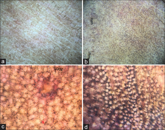
- Vinay K, Bishnoi A, Parsad D, Saikia UN, Sendhil Kumaran M. Dermoscopic evaluation and histopathological correlation of acquired dermal macular hyperpigmentation. Int J Dermatol. 2017;56:1395–9. - PubMed
-
- Chandran V, Kumarasinghe SP. Macular pigmentation of uncertain aetiology revisited: Two case reports and a proposed algorithm for clinical classification. Australas J Dermatol. 2017;58:45–9. - PubMed
-
- Ebihara T, Nakayama H. Pigmented contact dermatitis. Clin Dermatol. 1997;15:593–9. - PubMed
-
- Robles-Méndez JC, Rizo-Frías P, Herz-Ruelas ME, Pandya AG, Ocampo Candiani J. Lichen planus pigmentosus and its variants: Review and update. Int J Dermatol. 2018;57:505–14. - PubMed
-
- Zaynoun S, Rubeiz N, Kibbi A-G. Ashy dermatoses – A critical review of the literature and a proposed simplified clinical classification. Int J Dermatol. 2008;47:542–4. - PubMed
Publication types
LinkOut - more resources
Full Text Sources
