

Clinicopathological Profile of Childhood Onset Cutaneous Mastocytosis from a Tertiary Care Center in South India
- PMID: 34667757
- PMCID: PMC8456265
- DOI: 10.4103/idoj.IDOJ_924_20
Clinicopathological Profile of Childhood Onset Cutaneous Mastocytosis from a Tertiary Care Center in South India
Abstract
Background: Mastocytosis is characterized by clonal proliferation of mast cells in various organs and can have isolated cutaneous or systemic involvement. Childhood-onset mastocytosis (COM) is usually cutaneous and regresses spontaneously, while adult-onset mastocytosis (AOM) is often persistent with systemic involvement. There is limited data on COM from India.
Objective: To elucidate the clinicopathological profile of COM.
Methods: We conducted a retrospective chart review of all the patients with histologically proven COM (≤16 years), presenting over 11 years (January 2009 to December 2019) to the Dermatology Department. We compiled the demographic data, clinical characteristics (morphology, extent, distribution), laboratory investigations, histopathology findings, imaging (ultrasound abdomen), c-KIT mutation results, where available, and other associated abnormalities, and grouped them according to the WHO classification for mastocytosis.
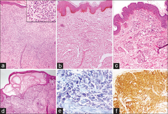
Results: Among the 66 patients with COM (M: F-1.6:1), 89.4% had onset before 2 years of age. The subtypes were: maculopapular cutaneous mastocytosis (MPCM: 44, 66.7%); mastocytoma of the skin (MOS: 19, 28.8%); diffuse cutaneous mastocytosis (DCM: 2, 3%) and indolent systemic mastocytosis (ISM: 1, 1.5%). Blistering was observed in 29 (43.9%) and Darier sign was elicited in 47 (71.2%) patients. Serum tryptase was elevated in 9/21 (42.9%) patients, but none had systemic mastocytosis. Three patients had c-KIT mutations (two in exon 8 and one in exon 17). Most patients were managed symptomatically and the patient with ISM improved with imatinib.
Conclusion: MPCM is the most common variant of COM and most patients had a disease onset before 2 years. Overall, COM had a good prognosis with rare systemic involvement, mitigating the need for extensive evaluation routinely in children.
Keywords: Childhood mastocytosis; c-KIT; cutaneous mastocytosis; mastocytoma; pediatric mastocytosis; tryptase; urticaria pigmentosa.
Copyright: © 2021 Indian Dermatology Online Journal.
Conflict of interest statement
There are no conflicts of interest.
Figures


Similar articles
-
Large maculopapular cutaneous lesions are associated with favorable outcome in childhood-onset mastocytosis.J Allergy Clin Immunol. 2015 Dec;136(6):1581-1590.e3. doi: 10.1016/j.jaci.2015.05.034. Epub 2015 Jul 4. J Allergy Clin Immunol. 2015. PMID: 26152315
-
Current Challenges in the Diagnosis of Pediatric Cutaneous Mastocytosis.Diagnostics (Basel). 2023 Dec 1;13(23):3583. doi: 10.3390/diagnostics13233583. Diagnostics (Basel). 2023. PMID: 38066824 Free PMC article. Review.
-
Mastocytosis in children: a single-center long-term follow-up study.Int J Dermatol. 2023 May;62(5):616-620. doi: 10.1111/ijd.16612. Epub 2023 Feb 20. Int J Dermatol. 2023. PMID: 36807903
-
Cutaneous mastocytosis in childhood.Allergol Select. 2022 Jan 5;6:1-10. doi: 10.5414/ALX02304E. eCollection 2022. Allergol Select. 2022. PMID: 35028497 Free PMC article. Review.
-
Solitary mastocytoma presenting in an adult: report and literature review of adult-onset solitary cutaneous mastocytoma with recommendations for evaluation and treatment.Dermatol Pract Concept. 2016 Jul 31;6(3):31-8. doi: 10.5826/dpc.0603a07. eCollection 2016 Jul. Dermatol Pract Concept. 2016. PMID: 27648381 Free PMC article. Review.
Cited by
-
Characteristics and Therapeutic Strategies for Diffuse Cutaneous Mastocytosis.JAMA Dermatol. 2025 Aug 1;161(8):855-862. doi: 10.1001/jamadermatol.2025.1488. JAMA Dermatol. 2025. PMID: 40434754
References
-
- Méni C, Bruneau J, Georgin-Lavialle S, Le Saché de Peufeilhoux L, Damaj G, Hadj-Rabia S, et al. Pediatric mastocytosis: A systematic review of 1747 cases. Br J Dermatol. 2015;172:642–51. - PubMed
-
- Hartmann K, Escribano L, Grattan C, Brockow K, Carter MC, Alvarez-Twose I, et al. Cutaneous manifestations in patients with mastocytosis: Consensus report of the European Competence Network on Mastocytosis; the American Academy of Allergy, Asthma and Immunology; and the European Academy of Allergology and Clinical Immunology. J Allergy Clin Immunol. 2016;137:35–45. - PubMed
-
- Bodemer C, Hermine O, Palmérini F, Yang Y, Grandpeix-Guyodo C, Leventhal PS, et al. Pediatric mastocytosis is a clonal disease associated with D816V and other activating c-KIT mutations. J Invest Dermatol. 2010;130:804–15. - PubMed
-
- Azaña JM, Torrelo A, Matito A. Update on mastocytosis (part 1): Pathophysiology, clinical features, and diagnosis. Actas Dermosifiliogr. 2016;107:5–14. - PubMed
LinkOut - more resources
Full Text Sources
