

doi: 10.15252/embr.202154046.
Epub 2021 Oct 20.
AI revolutions in biology: The joys and perils of AlphaFold
Affiliations
- PMID: 34668287
- PMCID: PMC8567224
- DOI: 10.15252/embr.202154046
Item in Clipboard
AI revolutions in biology: The joys and perils of AlphaFold
EMBO Rep.
.
Abstract
AlphaFold is the most ground-breaking application of AI in science so far; it will revolutionize structural biology, but caution is warranted.
© 2021 The Authors. Published under the terms of the CC BY 4.0 license.
Figures
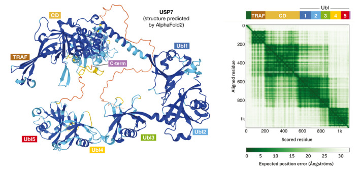
(Left) A ribbon model of USP7 exported directly from the EBI Web server; the coloring ranges from blue (high confidence) to red (low confidence); the domain names are annotated manually. (Right) The AlphaFold matrix showing the expected position error for each residue in the sequence; a detailed explanation of the matrix is available at the EBI Web server; the top bar with domain annotation and names was added manually (figures downloaded from the AlphaFold webserver).
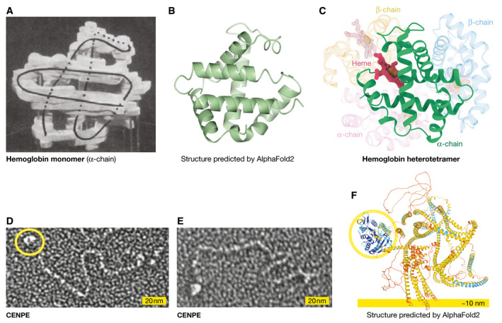
(A) The hemoglobin α‐chain monomer structure by Max Perutz and co‐workers; from Perutz et al (1960) Nature 185: 416–22, with permission from Springer Nature; (B) the AlphaFold model, predicted correctly, but lacking ligands and partners, displayed in the same orientation (created by CCP4MG (McNicholas et al, 2011); (C) a diagram of the hemoglobin fold that is a heterotetramer of two α and two β chains, each containing a heme coordinating a Fe2+ ion. (D,E) an electron micrograph (kindly provided by Y. Kim and D Cleveland) of CENPE showing the motor domain (yellow circle) and the coiled‐coil region that extends several hundreds of Ångström; (F) the AlphaFold model showing the motor domain (yellow circle) that is predicted with high confidence, and the “warped” set of helices that are predicted with low confidence and do not agree with the experimental data (image downloaded from the AlphaFold Web server).
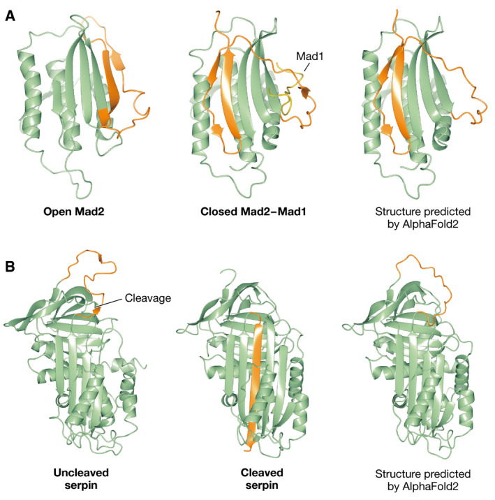
(A) The open Mad2 solution structure determined by NMR (left) undergoes a rearrangement of two beta strands (shown in orange), in which Mad2 embraces the Mad1 partner (shown as a yellow ribbon), a structure that was determined by X‐ray crystallography (middle); AlphaFold2 erroneously predicts the structure of the refolded complex with Mad1 as the native structure of Mad2 (right). (B) The structure of an uncleaved Serpin (left) with a long loop (shown in orange), which after proteolytic cleavage inserts as a β‐strand in the middle of the β‐sheet; AlphaFold correctly predicts the structure of the uncleaved protein. All figure panels were created by CCP4MG.
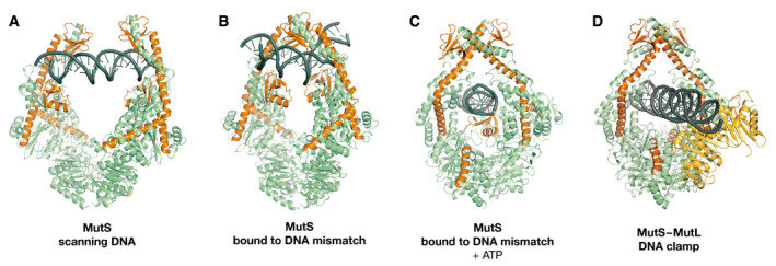
Structures of scanning MutS (A), mismatch‐bound MutS (B), transition‐state MutS (C), and MutLLN40‐bound MutS (D). Monomer A is shown in green, with monomer B in blue, MutLLN40 in orange, and DNA in black.
References
-
- Berman H, Henrick K, Nakamura H (2003) Announcing the worldwide protein data bank. Nat Struct Mol Biol 10: 980 - PubMed
-
- Evans R, O’Neill M, Pritzel A, Antropova N, Senior A, Green T, Žídek A, Bates R, Blackwell S, Yim J et al (2021) Protein complex prediction with AlphaFold‐Multimer. bioRxiv 10.1101/2021.10.04.463034 [PREPRINT] - DOI
Publication types
MeSH terms
LinkOut - more resources
Full Text Sources
Other Literature Sources
