

Acoustofluidic medium exchange for preparation of electrocompetent bacteria using channel wall trapping
- PMID: 34668506
- PMCID: PMC8577197
- DOI: 10.1039/d1lc00406a
Acoustofluidic medium exchange for preparation of electrocompetent bacteria using channel wall trapping
Abstract
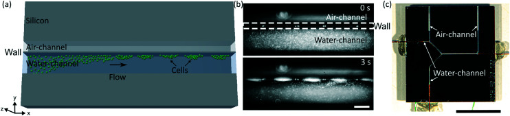
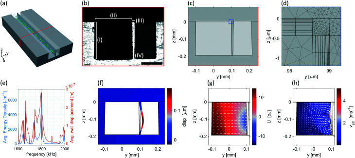
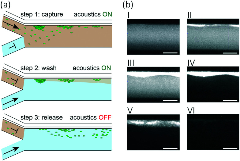
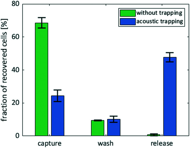
Comprehensive integration of process steps into a miniaturised version of synthetic biology workflows remains a crucial task in automating the design of biosystems. However, each of these process steps has specific demands with respect to the environmental conditions, including in particular the composition of the surrounding fluid, which makes integration cumbersome. As a case in point, transformation, i.e. reprogramming of bacteria by delivering exogenous genetic material (such as DNA) into the cytoplasm, is a key process in molecular engineering and modern biotechnology in general. Transformation is often performed by electroporation, i.e. creating pores in the membrane using electric shocks in a low conductivity environment. However, cell preparation for electroporation can be cumbersome as it requires the exchange of growth medium (high-conductivity) for low-conductivity medium, typically performed via multiple time-intensive centrifugation steps. To simplify and miniaturise this step, we developed an acoustofluidic device capable of trapping the bacterium Escherichia coli non-invasively for subsequent exchange of medium, which is challenging in acoustofluidic devices due to detrimental acoustic streaming effects. With an improved etching process, we were able to produce a thin wall between two microfluidic channels, which, upon excitation, can generate streaming fields that complement the acoustic radiation force and therefore can be utilised for trapping of bacteria. Our novel design robustly traps Escherichia coli at a flow rate of 10 μL min-1 and has a cell recovery performance of 47 ± 3% after washing the trapped cells. To verify that the performance of the medium exchange device is sufficient, we tested the electrocompetence of the recovered cells in a standard transformation procedure and found a transformation efficiency of 8 × 10⁵ CFU per μg of plasmid DNA. Our device is a low-volume alternative to centrifugation-based methods and opens the door for miniaturisation of a plethora of microbiological and molecular engineering protocols.
Conflict of interest statement
The authors declare no competing financial interest.
Figures
Similar articles
-
Scalable Device for Automated Microbial Electroporation in a Digital Microfluidic Platform.ACS Synth Biol. 2017 Sep 15;6(9):1701-1709. doi: 10.1021/acssynbio.7b00007. Epub 2017 Jun 7. ACS Synth Biol. 2017. PMID: 28569062
-
Optimization of electroporation-mediated transformation: Staphylococcus carnosus as model organism.J Appl Microbiol. 2007 Mar;102(3):736-47. doi: 10.1111/j.1365-2672.2006.03127.x. J Appl Microbiol. 2007. PMID: 17309623
-
Improved method for high-efficiency electrotransformation of Escherichia coli with the large BAC plasmids.Folia Microbiol (Praha). 2014 Jan;59(1):53-61. doi: 10.1007/s12223-013-0267-1. Epub 2013 Jul 12. Folia Microbiol (Praha). 2014. PMID: 23846555
-
Electric field-induced effects on neuronal cell biology accompanying dielectrophoretic trapping.Adv Anat Embryol Cell Biol. 2003;173:III-IX, 1-77. doi: 10.1007/978-3-642-55469-8. Adv Anat Embryol Cell Biol. 2003. PMID: 12901336 Review.
-
Optimization of electrotransformation (ETF) conditions in lactic acid bacteria (LAB).J Microbiol Methods. 2020 Jul;174:105944. doi: 10.1016/j.mimet.2020.105944. Epub 2020 May 15. J Microbiol Methods. 2020. PMID: 32417130 Review.
Cited by
-
Harnessing the power of Microscale AcoustoFluidics: A perspective based on BAW cancer diagnostics.Biomicrofluidics. 2024 Feb 29;18(1):011304. doi: 10.1063/5.0180158. eCollection 2024 Jan. Biomicrofluidics. 2024. PMID: 38434238 Free PMC article.
-
Biomolecular actuators for genetically selective acoustic manipulation of cells.Sci Adv. 2023 Feb 22;9(8):eadd9186. doi: 10.1126/sciadv.add9186. Epub 2023 Feb 22. Sci Adv. 2023. PMID: 36812320 Free PMC article.
-
Development of a Mass-Producible Microfluidic Device for Single and Bulk Mycobacteria Investigations.Biosensors (Basel). 2025 Feb 13;15(2):108. doi: 10.3390/bios15020108. Biosensors (Basel). 2025. PMID: 39997010 Free PMC article.
References
Publication types
MeSH terms
Substances
LinkOut - more resources
Full Text Sources
Research Materials
Miscellaneous