. 2022 Feb 9;96(3):e0109821.
doi: 10.1128/JVI.01098-21. Epub 2021 Oct 20.

Evolution and Diversity of Bat and Rodent Paramyxoviruses from North America

Affiliations

Evolution and Diversity of Bat and Rodent Paramyxoviruses from North America

Brendan B Larsen et al. J Virol. .

Abstract

Paramyxoviruses are a diverse group of negative-sense, single-stranded RNA viruses of which several species cause significant mortality and morbidity. In recent years the collection of paramyxovirus sequences detected in wild mammals has substantially grown; however, little is known about paramyxovirus diversity in North American mammals. To better understand natural paramyxovirus diversity, host range, and host specificity, we sought to comprehensively characterize paramyxoviruses across a range of diverse cooccurring wild small mammals in southern Arizona. We used highly degenerate primers to screen fecal and urine samples and obtained a total of 55 paramyxovirus sequences from 12 rodent species and 6 bat species. We also performed Illumina transcriptome sequencing (RNA-seq) and de novo assembly on 14 of the positive samples to recover a total of 5 near-full-length viral genomes. We show there are at least two clades of rodent-borne paramyxoviruses in Arizona, while bat-associated paramyxoviruses formed a putative single clade. Using structural homology modeling of the viral attachment protein, we infer that three of the five novel viruses likely bind sialic acid in a manner similar to other respiroviruses, while the other two viruses from heteromyid rodents likely bind a novel host receptor. We find no evidence for cross-species transmission, even among closely related sympatric host species. Taken together, these data suggest paramyxoviruses are a common viral infection in some bat and rodent species present in North America and illuminate the evolution of these viruses. IMPORTANCE There are a number of viral lineages that are potential zoonotic threats to humans. One of these, paramyxoviruses have jumped into humans multiple times from wild and domestic animals. We conducted one of the largest viral surveys of wild mammals in the United States to better understand paramyxovirus diversity and evolution.

Keywords: Paramyxovirus; codivergence; evolution; mammals.


Figures

FIG 1
FIG 1
Elevation relief map showing sampling locations across southeast Arizona. Each sampling location is represented with a red dot and a 1-letter code which corresponds to the information in Table S1 with capture information for all species. For each location, positive species are shown with their 4-letter species code.
FIG 2
FIG 2
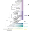
Maximum likelihood phylogeny from the concatenated RMH (∼490 nt) and PAR (∼550 nt) sequences with reference paramyxoviruses (gray) and novel paramyxoviruses found in this study (bold). Host species are included in the name for each viral sequence generated in this study. Recognized or proposed genera within Orthoparamyxovirinae are colored and named. Host identity is shown next to each clade we sampled a virus of in our study. The tree was midpoint rooted.
FIG 3
FIG 3
(A and B)Maximum likelihood phylogenies of (A) concatenated M and L sequences or (B) near-full-length L. Reference paramyxoviruses are shown in italics, with sequences from this study in bold. For sequences from this study, the host species is given in the name. Taxa that moved clade placement based on new sequence information from Fig. 2 are shown in red. Bootstrap support is shown by each node. The trees were midpoint rooted.
FIG 4
FIG 4
Maximum likelihood phylogeny of RMH. Visualization is the same as Fig. 2.
FIG 5
FIG 5
Maximum likelihood gene trees built from nucleotide sequences for four genes from representative paramyxoviruses for which complete genome sequences are available (in bold, from this study). The established genera Respirovirus, Henipavirus, Morbillivirus, and Narmovirus are colored, while Jeilongvirus is not colored since it does not form a single clade in the four genes. Only bootstrap values of <0.85 are shown. For each gene tree, sequences were clipped with Gblocks, and the 3rd position in codon was stripped. The trees were midpoint rooted.
FIG 6
FIG 6
Genome structure with putative genes based on ORF homology of reference paramyxoviruses (gray) with novel viruses from this study (in bold). The phylogeny on the right is based on amino acid alignments of complete L genes that were clipped using G-blocks to only include amino acid positions where homology could be reliably assessed. The phylogeny is only meant to show the putative relationships of the viruses to understand the gain and loss of different genes. Branch lengths are not to scale, and no node support was included. Question marks are used on the RBP locus if the gene does not contain the canonical sialic acid active site and has a putative unknown receptor. The BA5 assembly did not include the most 3′ end of the genome and so is missing.
FIG 7
FIG 7
Comparison of key amino acids in the active site of sialic acid binding viruses at the HN RBP. (A) The positions of the binding site amino acid residues and numbering of human parainfluenza-3 (now human respirovirus 3), PDB:1V3D, in red. Residues in light blue are the inferred binding site of BC5 (from the bat species Tadarida brasiliensis) based on structural modeling. In black is the sialic acid ligand N-acetylneuraminic acid, and blue lines represent inferred hydrogen bonds between residues and the sialic acid. (B) The same active site amino acid residues involved in binding for different representative paramyxoviruses (light gray) with the novel paramyxoviruses from this study (in bold, with host species included in name). Also included is the neuraminidase hexapeptide conserved sequence (amino acid [AA] positions 252 to 257). Amino acid differences from the human respirovirus 3 sequence are colored. The phylogeny on the right is the same as in Fig. 6.
FIG 8
FIG 8
Maximum likelihood RMH phylogeny showing the relationship of P. maniculatus and P. leucopus paramyxoviruses sampled in a single high-density area. The tree is rooted with the closest Peromyscus paramyxovirus we sampled (sample CH10, from Peromyscus boylii).
FIG 9
FIG 9
Maximum likelihood phylogenies showing relationships between host (left) and virus (right) of putative Shaanvirus members. Bat families are shaded.

References

    1. Jones KE, Patel NG, Levy MA, Storeygard A, Balk D, Gittleman JL, Daszak P. 2008. Global trends in emerging infectious diseases. Nature 451:990–993. 10.1038/nature06536. - DOI - PMC - PubMed
    1. Taylor LH, Latham SM, Woolhouse MEJ. 2001. Risk factors for human disease emergence. Philos Trans R Soc Lond B Biol Sci 356:983–989. 10.1098/rstb.2001.0888. - DOI - PMC - PubMed
    1. Morens DM, Folkers GK, Fauci AS. 2008. Emerging infections: a perpetual challenge. Lancet Infect Dis 8:710–719. 10.1016/S1473-3099(08)70256-1. - DOI - PMC - PubMed
    1. Zhou P, Yang X-L, Wang X-G, Hu B, Zhang L, Zhang W, Si H-R, Zhu Y, Li B, Huang C-L, Chen H-D, Chen J, Luo Y, Guo H, Jiang R-D, Liu M-Q, Chen Y, Shen X-R, Wang X, Zheng X-S, Zhao K, Chen Q-J, Deng F, Liu L-L, Yan B, Zhan F-X, Wang Y-Y, Xiao G-F, Shi Z-L. 2020. A pneumonia outbreak associated with a new coronavirus of probable bat origin. Nature 579:270–273. 10.1038/s41586-020-2012-7. - DOI - PMC - PubMed
    1. Andersen KG, Rambaut A, Lipkin WI, Holmes EC, Garry RF. 2020. The proximal origin of SARS-CoV-2. Nat Med 26:450–452. 10.1038/s41591-020-0820-9. - DOI - PMC - PubMed

Publication types

MeSH terms

LinkOut - more resources