. 2022 Jan 12;96(1):e0149221.
doi: 10.1128/JVI.01492-21. Epub 2021 Oct 20.

Susceptibilities of Human ACE2 Genetic Variants in Coronavirus Infection

Affiliations

Susceptibilities of Human ACE2 Genetic Variants in Coronavirus Infection

Wenlin Ren et al. J Virol. .

Abstract

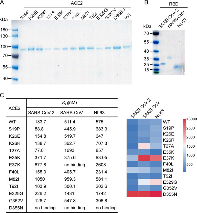
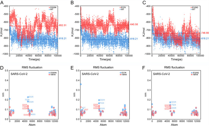
The coronavirus disease 2019 (COVID-19) pandemic, caused by severe acute respiratory syndrome coronavirus 2 (SARS-CoV-2), has resulted in more than 235 million cases worldwide and 4.8 million deaths (October 2021), with various incidences and mortalities among regions/ethnicities. The coronaviruses SARS-CoV, SARS-CoV-2, and HCoV-NL63 utilize the angiotensin-converting enzyme 2 (ACE2) as the receptor to enter cells. We hypothesized that the genetic variability in ACE2 may contribute to the variable clinical outcomes of COVID-19. To test this hypothesis, we first conducted an in silico investigation of single-nucleotide polymorphisms (SNPs) in the coding region of ACE2. We then applied an integrated approach of genetics, biochemistry, and virology to explore the capacity of select ACE2 variants to bind coronavirus spike proteins and mediate viral entry. We identified the ACE2 D355N variant that restricts the spike protein-ACE2 interaction and consequently limits infection both in vitro and in vivo. In conclusion, ACE2 polymorphisms could modulate susceptibility to SARS-CoV-2, which may lead to variable disease severity. IMPORTANCE There is considerable variation in disease severity among patients infected with SARS-CoV-2, the virus that causes COVID-19. Human genetic variation can affect disease outcome, and the coronaviruses SARS-CoV, SARS-CoV-2, and HCoV-NL63 utilize human ACE2 as the receptor to enter cells. We found that several missense ACE2 single-nucleotide variants (SNVs) that showed significantly altered binding with the spike proteins of SARS-CoV, SARS-CoV-2, and NL63-HCoV. We identified an ACE2 SNP, D355N, that restricts the spike protein-ACE2 interaction and consequently has the potential to protect individuals against SARS-CoV-2 infection. Our study highlights that ACE2 polymorphisms could impact human susceptibility to SARS-CoV-2, which may contribute to ethnic and geographical differences in SARS-CoV-2 spread and pathogenicity.

Keywords: ACE2; COVID-19; HCoV-NL63; SARS-CoV; SARS-CoV-2; SNP.


Figures

FIG 1
FIG 1
Schematic representation of the ACE2 molecule and positions of the studied SNP loci. (A) Structures of human ACE2 complexed with the spike proteins of SARS-CoV-2 (PDB code 6M0J), SARS-CoV (PDB code 2AJF), or HCoV-NL63 (PDB code 3KBH). ACE2 and the spike protein of each virus are colored in green and cyan, respectively. The residues of ACE2 at the interface with each spike protein are highlighted. (B) Coding region variants from gnomAD in the genes encoding ACE2 used in this study are indicated. The SNPs and the alteration of the amino acids in this study are shown. (C) Prediction of the interaction of coronavirus spike proteins with ACE2 variants. The ΔΔG for missense mutation was calculated by mCSM-PPI2 with PBD codes 2AJF (SARS-CoV spike complexed with human ACE2), 6M0J (SARS-CoV-2 spike complexed with human ACE2), or 3KBH (NL63-CoV spike complexed with human ACE2) as the model. The individual SNPs are named according to their identification numbers registered at the SNP database (dbSNP). The allele frequency of SNPs are as referenced in gnomAD database.
FIG 2
FIG 2
ACE2 variants bind viral spike proteins. (A) HeLa cells transduced with ACE2 variants were incubated with the recombinant S1 domain of the SARS-CoV-2, SARS-CoV, or HCoV-NL63 spike proteins C-terminally fused with Fc (1 μg/ml). Cells were then incubated with goat anti-human IgG (H+L) conjugated to Alexa Fluor 647 followed by flow cytometry analysis. Values are binding efficiencies defined as the percentage of ACE2-expressing cells (zsGreen1+) positive for S1-Fc. Values are means plus standard deviations (SD) (error bars) (n = 3). This experiment was independently repeated three times with similar results. (B) HeLa cells transduced with lentiviruses expressing FLAG-tagged human ACE2 variants were subjected to immunoblotting. Tubulin served as the loading control. This experiment was independently repeated three times with similar results. A representative blot is shown. (C) HeLa cells transduced with lentiviruses (pLVX-IRES-zsGreen1) expressing ACE2 variants as indicated were incubated with rabbit polyclonal antibody (Sino Biological Inc. China; catalog no. 10108-T24) against ACE2. The cells were washed and then stained with 2 μg/ml goat anti-rabbit IgG (H+L) conjugated with Alexa Fluor 568 and DAPI (1 μg/ml). The cell images were captured with a Zeiss LSM 880 confocal microscope. ACE2 on cell surface was shown in the merge images processed by ZEN3.2 software. This experiment was independently repeated three times with similar results, and the representative images are shown. (D) HeLa cells transduced with ACE2 variants were incubated with increasing doses (1 μg/ml, 5 μg/ml, or 10 μg/ml) of the recombinant S1 domain of SARS-CoV-2 (left), SARS-CoV (middle), or HCoV-NL63 (right) spike proteins fused to Fc. The other procedure and binding efficiency were performed as described in panel A. Values are means plus standard deviations (SD) (error bars) (n = 3). This experiment was independently repeated three times with similar results.
FIG 3
FIG 3
SPR analysis of ACE2 variants binding viral spike proteins. (A and B) The N-terminal peptidase domain of each human ACE2 (residues Met1-Asp615) variant (A), SARS-CoV-2 RBD (residues Thr333-Pro527), SARS-CoV RBD (residues Arg306-Leu515), or NL63-CoV RBD (residues Gln481-Ile616) (B) were expressed and purified as described in Materials and Methods. The purified proteins were analyzed by SDS-PAGE with Coomassie blue staining. (C) Affinity of ACE2 SNVs for coronavirus spike proteins. Dissociation constant (Kd) values were determined by SPR and are presented as a heatmap according to the indicated color legend. This experiment was independently repeated three times with similar results.
FIG 4
FIG 4
MD simulation results of the wild type and the mutants for different ACE2-RBDs complexes. (A to C) Nonbonded interaction energies between RBD and different mutants as follows for D355N (A), E37K (B), and E329G (C). The blue and red represent interaction energies between ACE2-RBD complexes of wild-type and mutants. Each point represents interaction energy between ACE2 and RBD at a certain time, whereas lines represent the average energies. (D to F) Cα-RMSFs of interface atoms for D355N (D), E37K (E), and E329G (F).
FIG 5
FIG 5
The ability of ACE2 variants to facilitate entry of virus pseudotyped with coronavirus spike proteins. (A and B) HeLa cells transduced with lentiviruses expressing ACE2 orthologs, SNVs, or empty vector were infected with virus pseudotyped with SARS-CoV spike, SARS-CoV-2 spike, or VSV-G and containing a firefly luciferase (Fluc) reporter gene. Intracellular Fluc activity of cell lysates was determined after 48 h of infection. Values are means plus standard deviations (SD) (error bars) (n = 3). This experiment was independently repeated at least three times. **, P < 0.01; ***, P < 0.001. Significance assessed by one-way ANOVA.
FIG 6
FIG 6
The ACE2 variants mediate authentic SARS-CoV-2 virus infection in vitro. (A) HeLa cells transduced with lentiviruses expressing human ACE2 SNVs or mouse ACE2 were infected with various doses of SARS-CoV-2 virus (MOI, 1, 0.3, 0.1, or 0.03). Expression of the viral nucleocapsid (N) protein or ACE2 orthologs was visualized by using the Operetta high content imaging system (PerkinElmer). Viral N protein (red) and ACE2 variant/ortholog (green) are shown. Scale bar, 100 μm. This experiment was independently repeated three times with similar results, and representative images are shown. (B) Images were analyzed and quantified using PerkinElmer Harmony high-content analysis software 4.9. The infection efficiency represents the percentage of SARS-CoV-2 infected cells/ACE2 positive cells (y axis). Values are means plus standard deviations (SD) (error bars) (n = 4). ns, no significance; **, P < 0.01. Significance assessed by one-way ANOVA.
FIG 7
FIG 7
Ability of ACE2 variants to mediate authentic virus entry in vivo. (A) Schematic representation of the experimental timeline. Wild-type BALB/c mice were transduced with empty recombinant adenovirus (Vector), or recombinant adenovirus expressing wild-type human ACE2 or the D355N variant for 3 days, followed by SARS-CoV-2 challenge. Mice were sacrificed at day 3 postinfection (n = 5 mice per group), and lung tissues were collected for immunostaining with anti-N serum (B) and viral load titration (C). Representative images are shown from n = 5 mice. Scale bar, 200 μm in panel B. Viral load was determined by focus-forming assay. **, P < 0.01. Significance assessed by one-way ANOVA in panel C.

References

    1. Fung TS, Liu DX. 2019. Human coronavirus: host-pathogen interaction. Annu Rev Microbiol 73:529–557. 10.1146/annurev-micro-020518-115759. - DOI - PubMed
    1. Lai MM. 1990. Coronavirus: organization, replication and expression of genome. Annu Rev Microbiol 44:303–333. 10.1146/annurev.mi.44.100190.001511. - DOI - PubMed
    1. Ksiazek TG, Erdman D, Goldsmith CS, Zaki SR, Peret T, Emery S, Tong S, Urbani C, Comer JA, Lim W, Rollin PE, Dowell SF, Ling AE, Humphrey CD, Shieh WJ, Guarner J, Paddock CD, Rota P, Fields B, DeRisi J, Yang JY, Cox N, Hughes JM, LeDuc JW, Bellini WJ, Anderson LJ, SARS Working Group . 2003. A novel coronavirus associated with severe acute respiratory syndrome. N Engl J Med 348:1953–1966. 10.1056/NEJMoa030781. - DOI - PubMed
    1. Zaki AM, van Boheemen S, Bestebroer TM, Osterhaus AD, Fouchier RA. 2012. Isolation of a novel coronavirus from a man with pneumonia in Saudi Arabia. N Engl J Med 367:1814–1820. 10.1056/NEJMoa1211721. - DOI - PubMed
    1. Morens DM, Fauci AS. 2020. Emerging pandemic diseases: how we got to COVID-19. Cell 182:1077–1092. 10.1016/j.cell.2020.08.021. - DOI - PMC - PubMed

Publication types

MeSH terms

Substances

LinkOut - more resources