. 2021 Nov;41(8):1709-1722.
doi: 10.1007/s10875-021-01133-0. Epub 2021 Oct 20.

SARS-CoV-2 Vaccine Induced Atypical Immune Responses in Antibody Defects: Everybody Does their Best

Affiliations

SARS-CoV-2 Vaccine Induced Atypical Immune Responses in Antibody Defects: Everybody Does their Best

Ane Fernandez Salinas et al. J Clin Immunol. 2021 Nov.

Abstract

Background: Data on immune responses to SARS-CoV-2 in patients with Primary Antibody Deficiencies (PAD) are limited to infected patients and to heterogeneous cohorts after immunization.

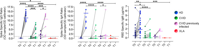
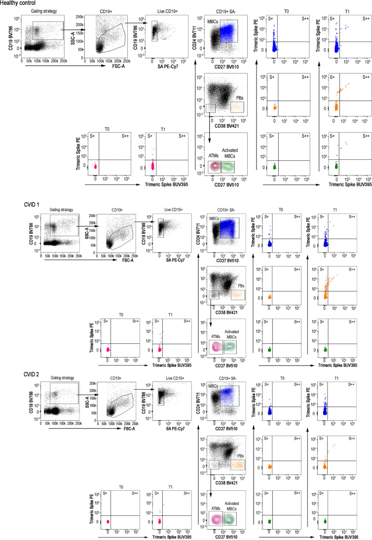
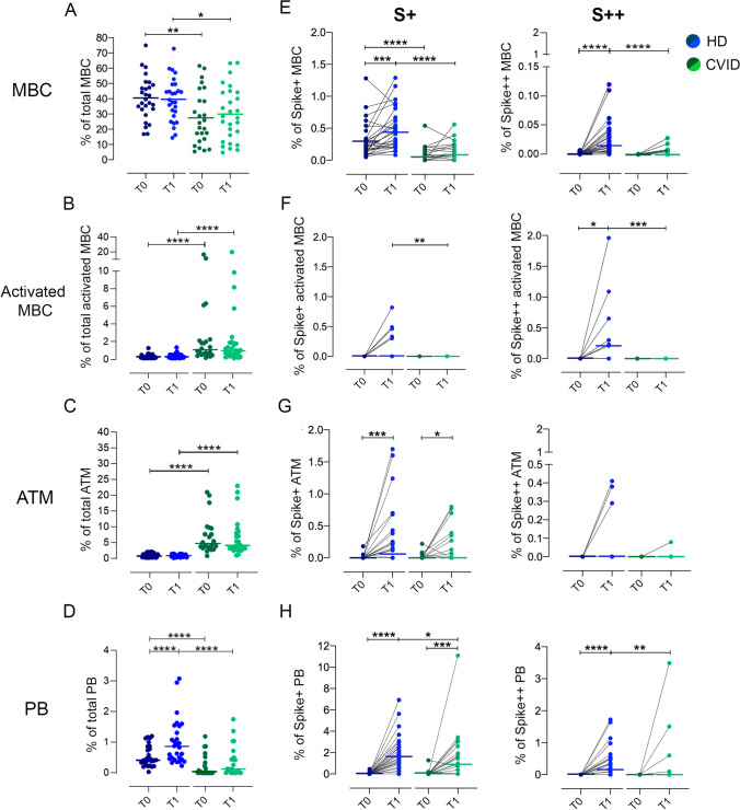
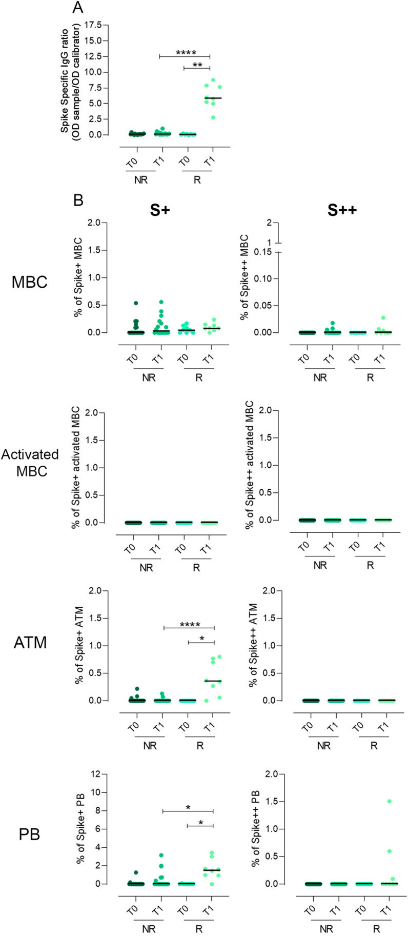
Methods: Forty-one patients with Common Variable Immune Deficiencies (CVID), six patients with X-linked Agammaglobulinemia (XLA), and 28 healthy age-matched controls (HD) were analyzed for anti-Spike and anti-receptor binding domain (RBD) antibody production, generation of Spike-specific memory B-cells, and Spike-specific T-cells before vaccination and one week after the second dose of BNT162b2 vaccine.

Results: The vaccine induced Spike-specific IgG and IgA antibody responses in all HD and in 20% of SARS-CoV-2 naive CVID patients. Anti-Spike IgG were detectable before vaccination in 4 out 7 CVID previously infected with SARS-CoV-2 and were boosted in six out of seven patients by the subsequent immunization raising higher levels than patients naïve to infection. While HD generated Spike-specific memory B-cells, and RBD-specific B-cells, CVID generated Spike-specific atypical B-cells, while RBD-specific B-cells were undetectable in all patients, indicating the incapability to generate this new specificity. Specific T-cell responses were evident in all HD and defective in 30% of CVID. All but one patient with XLA responded by specific T-cell only.

Conclusion: In PAD patients, early atypical immune responses after BNT162b2 immunization occurred, possibly by extra-follicular or incomplete germinal center reactions. If these responses to vaccination might result in a partial protection from infection or reinfection is now unknown. Our data suggests that SARS-CoV-2 infection more effectively primes the immune response than the immunization alone, possibly suggesting the need for a third vaccine dose for patients not previously infected.

Keywords: BNT162b2 vaccine; COVID-19; Common variable immune deficiencies; Memory cells; Primary antibody deficiencies; Receptor-binding-domain; SARS-CoV-2; Spike protein; X-linked agammaglobulinemia.


Conflict of interest statement

Authors declare that they have no conflict interest.

Figures

Fig. 1
Fig. 1
RBD-specific IgM and Spike-specific IgG and IgA antibodies in HD (blue circles), CVID patients (green circles), CVID previously infected patients (pink circles) and XLA (red circles), before (T0, dark color circles) and one week after the second dose (T1, lighter color circles) of BNT162b2 vaccine. For each group the median is shown as a bar. * P ≤ 0.05, ** P ≤  0.01, *** P ≤  0.001, **** P < 0.0001. Positive cut-off value is represented by a dashed line. N = 28 HD, N = 34 CVID, N = 6 XLA patients, and N = 7 CVID previously SARS-CoV-2 infected
Fig. 2
Fig. 2
Gating strategy to identify S + and S +  + MBCs, PBs, ATM , and activated MBCs. One HD and two CVID subjects are shown. We analyzed CD19 + B-cells, included in the live gate. SA PE-Cy7 was used as a decoy probe to gate out streptavidin-binding B-cells from further analysis. MBCs were identified as CD24 + CD27 + CD38-; PBs as CD24-CD27 +  + CD38 +  + ; ATMs as CD24-CD27-CD38-CD21- -; and activated MBCs as CD24- CD38-CD21-CD27 + . Flow cytometry plots show the staining patterns of SARS-CoV-2 antigen probes in the indicated B-cell populations during the follow-up. S + are B-cells that are Spike-PE + , but Spike-BUV395-. S +  + are instead Spike-PE + and SpikeBUV395 + . The color code identifies Spike + and +  + in MBCs (blue), PBs (orange), ATMs (dark red) ), and Activated MBCs (green)
Fig. 3
Fig. 3
Peripheral blood B-cells subsets, low and high binding capacity B-cells for recombinant Spike protein in HD (blue circles) and in CVID (green circles) before (T0, dark color circles) and after two doses of the BNT162b2 vaccine (T1, lighter color circles). We show the frequencies of peripheral blood MBCs (panel a), Activated MBCs (b), ATMs (c), and PBs (e) in HD (blue circles) and CVID (green circles). The frequency of S + and S +  +  B-cells inside each identified B-cell population is shown (e, f, g, and h). Medians are plotted as horizontal bars and statistical significance were determined using two-tailed Mann–Whitney U-test or Wilcoxon matched-pairs signed-rank test. *P < 0.05, **P < 0.01, ***P < 0.001; ****P < 0.0001. N = 28 HD and N = 26 CVID patients. B-cells subsets were defined as following: MBCs CD19 + CD24 + CD27 + CD38-; activated MBCs CD19 + , CD27 + CD24-CD38-; specific ATMs CD19 + CD27-CD24-CD28-; PBs CD19 + CD24-CD38 +  + CD27 +  +
Fig. 4
Fig. 4
Spike-specific IgG, low and high binding capacity B-cells for recombinant Spike protein in CVID who did not develop Spike-specific IgG (NR) and in CVID who developed Spike-specific IgG (R) before (T0) and after two doses of the BNT162b2 vaccine (T1). a Dot plot depicts the levels of Spike-specific IgG. b The frequency of S + and S +  +  B-cells inside each identified B-cell population, in R and NR CVID patients, is shown. Medians are plotted as horizontal bars and statistical significance were determined using two-tailed Mann–Whitney U-test or Wilcoxon matched-pairs signed-rank test. *P < 0.05, **P < 0.01, ****P < 0.0001. N = 16 NR and N = 8 R

References

    1. Chen G, Wu D, Guo W, Cao Y, Huang D, Wang H, et al. Clinical and immunological features of severe and moderate coronavirus disease 2019. J Clin Invest. 2020;130:2620–2629. - PMC - PubMed
    1. Picard C, Bobby Gaspar H, Al-Herz W, Bousfiha A, Casanova JL, Chatila T, et al. International union of immunological societies: 2017 primary immunodeficiency diseases committee report on inborn errors of immunity. J Clin Immunol. 2018;38:96–128. - PMC - PubMed
    1. Babaha F, Rezaei N. Primary immunodeficiency diseases in COVID-19 pandemic: a predisposing or protective factor? Am J Med Sci. 2020;360:740–741. - PMC - PubMed
    1. Meyts I, Bucciol G, Quinti I, Neven B, Fischer A, Seoane E, et al. Coronavirus disease 2019 in patients with inborn errors of immunity: An international study. J Allergy Clin Immunol. 2021;147:520–531. - PMC - PubMed
    1. Ho HE, Mathew S, Peluso MJ. Cunningham-rundles Ce. Clinical outcomes and features of COVID-19 in patients with primary immunodeficiencies in New York City. J Allergy Clin Immunol Pract. 2021;9:490–493. - PMC - PubMed

Publication types

MeSH terms