. 2022 Jan 11;88(1):e0095821.
doi: 10.1128/AEM.00958-21. Epub 2021 Oct 20.

Isolation and Characterization of Lignocellulose-Degrading Geobacillus thermoleovorans from Yellowstone National Park

Affiliations

Isolation and Characterization of Lignocellulose-Degrading Geobacillus thermoleovorans from Yellowstone National Park

Margaux M Meslé et al. Appl Environ Microbiol. .

Erratum in

Abstract

The microbial degradation of lignocellulose in natural ecosystems presents numerous biotechnological opportunities, including biofuel production from agricultural waste and feedstock biomass. To explore the degradation potential of specific thermophiles, we have identified and characterized extremophilic microorganisms isolated from hot springs environments that are capable of biodegrading lignin and cellulose substrates under thermoalkaline conditions, using a combination of culturing, genomics, and metabolomics techniques. Organisms that can use lignin and cellulose as a sole carbon source at 60 to 75°C were isolated from sediment slurry of thermoalkaline hot springs (71 to 81°C and pH 8 to 9) of Yellowstone National Park. Full-length 16S rRNA gene sequencing indicated that these isolates were closely related to Geobacillus thermoleovorans. Interestingly, most of these isolates demonstrated biofilm formation on lignin, a phenotype that is correlated with increased bioconversion. Assessment of metabolite level changes in two Geobacillus isolates from two representative springs were undertaken to characterize the metabolic responses associated with growth on glucose versus lignin carbon source as a function of pH and temperature. Overall, results from this study support that thermoalkaline springs harbor G. thermoleovorans microorganisms with lignocellulosic biomass degradation capabilities and potential downstream biotechnological applications. IMPORTANCE Since lignocellulosic biomass represents a major agro-industrial waste and renewable resource, its potential to replace nonrenewable petroleum-based products for energy production is considerable. Microbial ligninolytic and cellulolytic enzymes are of high interest in biorefineries for the valorization of lignocellulosic biomass, as they can withstand the extreme conditions (e.g., high temperature and high pH) required for processing. Of great interest is the ligninolytic potential of specific Geobacillus thermoleovorans isolates to function at a broad range of pH and temperatures, since lignin is the bottleneck in the bioprocessing of lignocellulose. In this study, results obtained from G. thermoleovorans isolates originating from YNP springs are significant because very few microorganisms from alkaline thermal environments have been discovered to have lignin- and cellulose-biodegrading capabilities, and this work opens new avenues for the biotechnological valorization of lignocellulosic biomass at an industrial scale.

Keywords: alkaline geothermal spring; biodegradation; biofuel; lignin; metabolomics.


Figures

FIG 1
FIG 1
Pictures of the thermoalkaline springs sampled in this study, located in the White Creek Drainage, Lower Geyser Basin, in Yellowstone National Park. The arrows indicate the sampling areas where spring water and sediment were collected.
FIG 2
FIG 2
Phylogenetic tree of the Geobacillus thermoleovorans isolates obtained by enrichment on lignin (L), cellulose (C), and xylose (X) substrates from Treefall (TF) and Five Sisters 2 (FS2) springs, based on Sanger sequencing of the 16S rRNA genes. The color coding on the tree branches represents the different substrates. C1 to C5 indicate colonies 1 to 5. The corresponding GenBank accession number for each isolate sequence is presented in Table 2. Only the isolate FS2 C C1 clustered with Brevibacillus thermoruber and was not used in the experiments performed in this study.
FIG 3
FIG 3
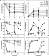
Headspace oxygen and carbon dioxide content (%) measured over time by GC in the lignin cultures containing G. thermoleovorans isolates from FS2 (squares) and TF (triangles) thermoalkaline springs, compared to the noninoculated controls (crosses). The assays were incubated with lignin (A), cellulose (B), and xylose (C) powders as the only carbon source (0.5 g/L, i.e., 5 mg initial content) at 75°C and pH 8, with air in the headspace. The dashed lines in the lignin assays represent two different isolates from each spring. Specific isolates (from Fig. 2) used in this experiment include: TF L C1 (full line), TF L C3 (dotted line), FS2 L C1 (full line), and another isolate (dotted line) from FS2 (lignin) that was not sequenced due to poor sequence quality; TF C C2 and FS2 C C5 (cellulose); and TF X C1 and FS2 X C1 (xylose). The arrows indicate the time of air exchange in the headspace. The error bars indicate the standard deviations for triplicate assays, and the injected air controls (dotted line and open circles) show the reproducibility of the GC measurements.
FIG 4
FIG 4
FE-SEM images obtained from the incubations at 70°C and pH 8.5 with lignin as the only carbon source. (A) Lignin matrix in the abiotic control (Mag = 49.13 KX, WD = 4.8 mm, EHT = 1.00 kV). (B) Formation of a biofilm on the lignin matrix by G. thermoleovorans cells of the TF L C1 isolate from Fig. 2 (Mag = 20.30 KX, WD = 5.1 mm, EHT = 1.00 kV). A clumping of the cells was observed visually in the culture tube pictured in Fig. S4.
FIG 5
FIG 5
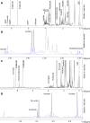
Representative 1D 1H NMR spectra of intracellular metabolite mixtures extracted from G. thermoleovorans cell cultures (isolate TF L C1, Fig. 2), grown aerobically at 70°C and pH 8.5 in minimal salt media with glucose (1 g/L) or lignin (1 g/L) as the sole carbon source, recorded on MSU’s 600-MHz solution NMR spectrometer. The x and y axes denote 1H chemical shift in ppm and relative signal intensity, respectively. Identified metabolites are labeled next to their characteristic signals with the position of the imidazole signal used as pH indicator. NMR signals assigned to specific signals are abbreviated as follows: DSS, sodium salt of 4,4-dimethyl-4-silapentane-1-sulfonic acid. (A and C) Representative 1D 1H NMR spectra of intracellular metabolites extracted from Geobacillus cell cultures grown on glucose (A) or lignin (C) as the sole carbon source. The data spans the full 1H chemical shift spectral range of 0 to ∼10 ppm. (B and D) Expanded spectral regions of the 1D 1H NMR spectra shown in panels A and C, respectively. In panels B and D, the original spectra are shown in gray, and identified metabolites are shown in blue as fitted signal intensities. Panel B depicts the spectral region spanning the 1.1- to 1.6-ppm 1H chemical shift range of the full spectrum shown in panel A, and panel D depicts the spectral region spanning the 1.9- to 2.7-ppm 1H chemical shift range of the full spectrum shown in panel C. All of the NMR chemical shifts and NMR signals are referenced to the DSS NMR signal set at 0 ppm.
FIG 6
FIG 6
Heat maps showing small molecule features measured via LC-MS in liquid cultures with the greatest power to differentiate cultures. (A) G. thermoleovorans isolates from TF (TF L C1; Fig. 2) and FS2 (FS2 L C1; Fig. 2) springs grown on lignin (0.5 g/L) at pH 8 and 70°C. Color gradient indicates relative concentration of the features, and m/z indicates their corresponding molecular weight (g/mol). The colored boxes highlight the main classes of compounds identified in the samples. (B) G. thermoleovorans from FS2 spring (isolate FS2 L C1, Fig. 2) grown on lignin (0.5 g/L) at pH 7 or 9 and at 60 or 70°C for 13 days.

References

    1. Arevalo-Gallegos A, Ahmad Z, Asgher M, Parra-Saldivar R, Iqbal HMN. 2017. Lignocellulose: a sustainable material to produce value-added products with a zero waste approach-A review. Int J Biol Macromol 99:308–318. 10.1016/j.ijbiomac.2017.02.097. - DOI - PubMed
    1. Gall DL, Ralph J, Donohue TJ, Noguera DR. 2017. Biochemical transformation of lignin for deriving valued commodities from lignocellulose. Curr Opin Biotechnol 45:120–126. 10.1016/j.copbio.2017.02.015. - DOI - PubMed
    1. Kim JS, Lee YY, Kim TH. 2016. A review on alkaline pretreatment technology for bioconversion of lignocellulosic biomass. Bioresour Technol 199:42–48. 10.1016/j.biortech.2015.08.085. - DOI - PubMed
    1. Anwar Z, Gulfraz M, Irshad M. 2014. Agro-industrial lignocellulosic biomass a key to unlock the future bio-energy: a brief review. J Radiat Res Applied Sci 7:163–173. 10.1016/j.jrras.2014.02.003. - DOI
    1. Beckham GT, Johnson CW, Karp EM, Salvachua D, Vardon DR. 2016. Opportunities and challenges in biological lignin valorization. Curr Opin Biotechnol 42:40–53. 10.1016/j.copbio.2016.02.030. - DOI - PubMed

Publication types

Supplementary concepts

LinkOut - more resources