Review
. 2022 Jan;23(1):23-34.
doi: 10.1038/s41583-021-00525-w. Epub 2021 Oct 20.

Emerging roles for CNS fibroblasts in health, injury and disease

Affiliations
Review

Emerging roles for CNS fibroblasts in health, injury and disease

Cayce E Dorrier et al. Nat Rev Neurosci. 2022 Jan.

Abstract

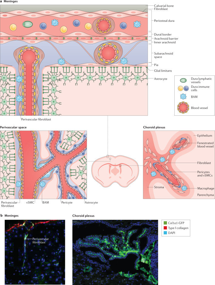
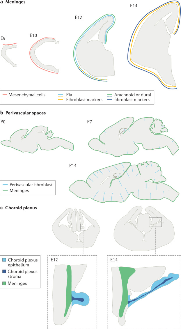
Recent transcriptomic, histological and functional studies have begun to shine light on the fibroblasts present in the meninges, choroid plexus and perivascular spaces of the brain and spinal cord. Although the origins and functions of CNS fibroblasts are still being described, it is clear that they represent a distinct cell population, or populations, that have likely been confused with other cell types on the basis of the expression of overlapping cellular markers. Recent work has revealed that fibroblasts play crucial roles in fibrotic scar formation in the CNS after injury and inflammation, which have also been attributed to other perivascular cell types such as pericytes and vascular smooth muscle cells. In this Review, we describe the current knowledge of the location and identity of CNS perivascular cell types, with a particular focus on CNS fibroblasts, including their origin, subtypes, roles in health and disease, and future areas for study.


Conflict of interest statement

The authors declare no competing interests.

Figures

Fig. 1
Fig. 1. Localization of fibroblasts in the adult mouse brain.
a | Fibroblasts are present in the meninges, choroid plexus and perivascular spaces. Fibroblasts are present in all three meningeal layers, the pia mater, arachnoid mater and dura mater,,–,. Different immune cell populations and vasculature (barrier, non-barrier blood vasculature and lymphatic vessels) are distributed between the leptomeninges (that is, the pia and arachnoid) and dura mater,. Perivascular fibroblasts surround blood vessels in the dura, leptomeninges, penetrating arterioles and pre-capillary arterioles with ‘ensheathing’ pericytes but not capillaries,,,,. Fibroblasts are located in the stroma (the inner region of the choroid plexus), which is surrounded by the epithelium, adjacent to non-barrier blood vasculature and macrophages,,. b | In adult Col1a1-GFP mice, expression of GFP from the Col1a1 promoter is used to mark fibroblasts in the meninges and perivascular spaces, (left) and within the stroma of the choroid plexus (right). BAM, border-associated macrophage; GFP, green fluorescent protein; vSMC, vascular smooth muscle cell. Left image in part b adapted from ref., Springer Nature Limited. Right image in part b adapted from ref., Elsevier.
Fig. 2
Fig. 2. Development of fibroblasts in different regions of the CNS.
a | Meningeal fibroblasts in the mouse forebrain first appear from the neural crest as undifferentiated mesenchymal cells at approximately embryonic day 9 (E9) and fully cover the forebrain by E10 (ref.). Some layer-specific meningeal fibroblast markers first appear ventrally in the mouse forebrain at E12 (RALDH2 for arachnoid fibroblasts; the neurotrophin receptor p75 for pial fibroblasts) but are expressed over the entire forebrain by E14 (ref.). b | Perivascular fibroblasts in the mouse brain parenchymal vasculature are infrequent at postnatal day 0 (P0) and located not far from the meninges. Over the next 2 weeks of postnatal development, many more vessels have perivascular fibroblasts and the cells are present deeper in the brain. c | The choroid plexus stroma of the lateral ventricle contains fibroblasts and the blood vasculature is spatially continuous with the adjacent meninges and extends along with the growth of the choroid plexus epithelium,,.
Fig. 3
Fig. 3. Organization of the glial and fibrotic scars.
In a healthy spinal cord, perivascular fibroblasts and macrophages reside in perivascular spaces,. In experimental autoimmune encephalomyelitis (EAE), a model of multiple sclerosis, neuroinflammatory lesions form in the white matter. These lesions include infiltrating immune cells, such as T cells, and a scar consisting of nearby fibroblasts and reactive astrocytes,. In spinal cord injury (SCI), a scar also forms in the area of the injury. However, the core of the injury site consists of an inner fibrotic scar containing extracellular matrix proteins, activated fibroblasts, microglia, and macrophages and an outer glial scar consisting of reactive astrocytes,,.

Similar articles

Cited by

References

    1. Vanlandewijck M, et al. A molecular atlas of cell types and zonation in the brain vasculature. Nature. 2018;554:475–480. - PubMed
    1. Dani N, et al. A cellular and spatial map of the choroid plexus across brain ventricles and ages. Cell. 2021;184:3056–3074.e21. - PMC - PubMed
    1. Rustenhoven J, et al. Functional characterization of the dural sinuses as a neuroimmune interface. Cell. 2021;184:1000–1016.e27. - PMC - PubMed
    1. Dorrier CE, et al. CNS fibroblasts form a fibrotic scar in response to immune cell infiltration. Nat. Neurosci. 2021;24:234–244. - PMC - PubMed
    1. Liu J, et al. A human cell type similar to murine central nervous system perivascular fibroblasts. Exp. Cell Res. 2021;402:112576. - PubMed

Publication types

MeSH terms