. 2022 Jun;43(6):1495-1507.
doi: 10.1038/s41401-021-00781-7. Epub 2021 Oct 20.

Baicalein ameliorates ulcerative colitis by improving intestinal epithelial barrier via AhR/IL-22 pathway in ILC3s

Affiliations

Baicalein ameliorates ulcerative colitis by improving intestinal epithelial barrier via AhR/IL-22 pathway in ILC3s

Yan-Yang Li et al. Acta Pharmacol Sin. 2022 Jun.

Abstract

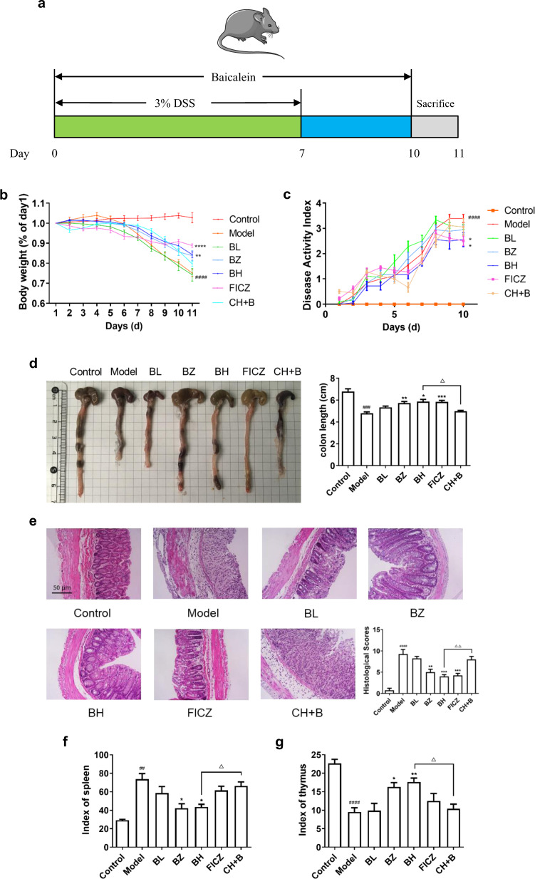
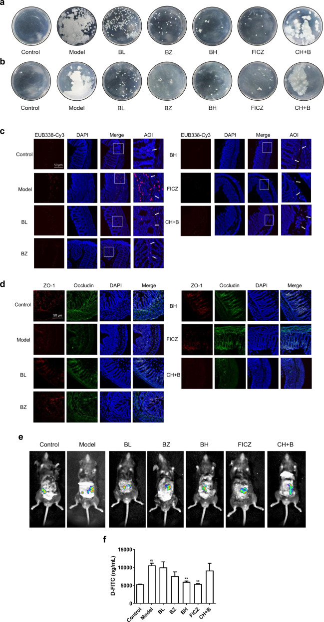
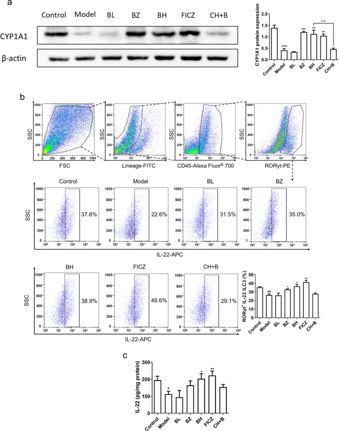
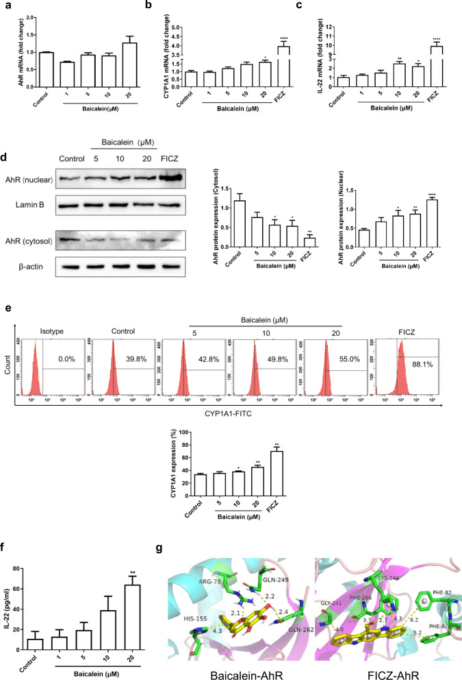
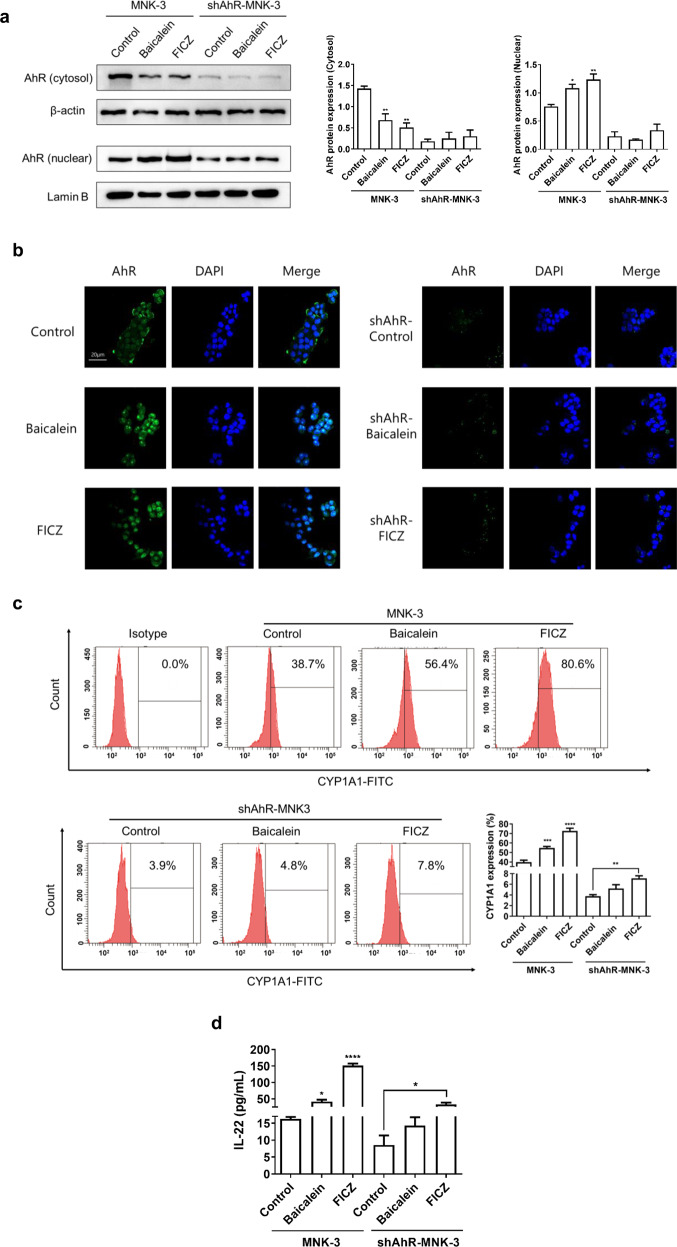
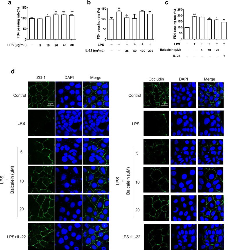
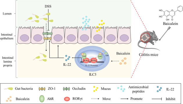
Ulcerative colitis (UC) is a chronic inflammatory disease of the gastrointestinal tract, which is closely related to gut barrier dysfunction. Emerging evidence shows that interleukin-22 (IL-22) derived from group 3 innate lymphoid cells (ILC3s) confers benefits on intestinal barrier, and IL-22 expression is controlled by aryl hydrocarbon receptor (AhR). Previous studies show that baicalein protects the colon from inflammatory damage. In this study we elucidated the molecular mechanisms underlying the protective effect of baicalein on intestinal barrier function in colitis mice. Mice were administered baicalein (10, 20, 40 mg·kg-1·d-1, i.g.) for 10 days; the mice freely drank 3% dextran sulfate sodium (DSS) on D1-D7 to induce colitis. We showed that baicalein administration simultaneously ameliorated gut inflammation, decreased intestinal permeability, restored tight junctions of colons possibly via promoting AhR/IL-22 pathway. Co-administration of AhR antagonist CH223191 (10 mg/kg, i.p.) partially blocked the therapeutic effects of baicalein in colitis mice, whereas AhR agonist FICZ (1 μg, i.p.) ameliorated symptoms and gut barrier function in colitis mice. In a murine lymphocyte line MNK-3, baicalein (5-20 μM) dose-dependently increased the expression of AhR downstream target protein CYP1A1, and enhanced IL-22 production through facilitating AhR nuclear translocation, these effects were greatly diminished in shAhR-MNK3 cells, suggesting that baicalein induced IL-22 production in AhR-dependent manner. To further clarify that, we constructed an in vitro system consisting of MNK-3 and Caco-2 cells, in which MNK-3 cell supernatant treated with baicalein could decrease FITC-dextran permeability and promoted the expression of tight junction proteins ZO-1 and occluding in Caco-2 cells. In conclusion, this study demonstrates that baicalein ameliorates colitis by improving intestinal epithelial barrier via AhR/IL-22 pathway in ILC3s, thus providing a potential therapy for UC.

Keywords: CH223191; FICZ; aryl hydrocarbon receptor; baicalein; epithelial barrier; group 3 innate lymphoid cells; interleukin-22; ulcerative colitis.


Conflict of interest statement

The authors declare no competing interests.

Figures

Fig. 1
Fig. 1. Baicalein ameliorated DSS-induced mice and the effect was dependent on AhR.
a Establishment of murine colitis model and administration of baicalein. b Body weight change. c DAI scores. d Length of colons. e Haematoxylin and eosin staining of colon (200×). f Index of spleen. g Index of thymus, n = 6 in each group. BL, low dose of baicalein (10 mg/kg); BZ, medium dose of baicalein (20 mg/kg); BH, high dose of baicalein (40 mg/kg); FICZ, AhR agonist; CH + B, CH223191 (AhR antagonist) + baicalein (40 mg/kg). #P < 0.05, ###P < 0.001, ####P < 0.001 vs. the control. *P < 0.05, **P < 0.01, ***P < 0.001 vs. the model. P < 0.05, △△P < 0.01 vs. the BH.
Fig. 2
Fig. 2. Baicalein improved the intestinal barrier structure and function in colitis mice.
a Bacterial loads in spleen were detected by bacteria culture. b Bacterial loads in MLNs were detected by bacteria culture. c Bacterial infiltration in intestine was observed using FISH (200×). d The expression levels of tight junction proteins ZO-1 and occludin were observed by immunofluorescence (200×). e FITC-dextran distribution in colitis mice was observed by small animal imaging. f Content of FITC-dextran in serum (n = 5). BL, low dose of baicalein (10 mg/kg); BZ, medium dose of baicalein (20 mg/kg); BH, high dose of baicalein (40 mg/kg); FICZ, AhR agonist; CH + B, CH223191 (AhR antagonist) + baicalein (40 mg/kg). ##P < 0.01 vs. the control. **P < 0.01, vs. the model.
Fig. 3
Fig. 3. Baicalein regulated AhR/IL-22 pathway in ILC3s of colitis mice.
a CYP1A1 protein expression of colon was evaluated using Western blot (n = 3). b The proportion of ILC3s was evaluated using flow cytometry (n = 6). c IL-22 content in colon was evaluated using ELISA (n = 5). BL, low dose of baicalein (10 mg/kg); BZ, medium dose of baicalein (20 mg/kg); BH, high dose of baicalein (40 mg/kg); FICZ, AhR agonist; CH + B, CH223191 (AhR antagonist) + baicalein (40 mg/kg). #P < 0.05, ##P < 0.01, ####P < 0.001 vs. the control. *P < 0.05, **P < 0.01, ***P < 0.001 vs. the model. △△P < 0.01 vs. the BH.
Fig. 4
Fig. 4. Baicalein activated AhR/IL-22 pathway in MNK-3 cells in vitro.
a AhR mRNA expression. b CYP1A1 mRNA expression. c IL-22 mRNA expression. d AhR protein expression in the cytoplasm and nucleus was detected using Western blot. e CYP1A1 protein expression was detected using flow cytometry; f The content of IL-22 secreted by MNK-3 cells was evaluated using ELISA. g Molecular docking models of baicalein-AhR and FICZ-AhR were constructed using AutoDock 1.5.6. n = 3 in each group. *P < 0.05, **P < 0.01, ****P < 0.001 vs. the control.
Fig. 5
Fig. 5. Establishment of shAhR-MNK3 stable transfected cell line.
a Transfection conditions for MNK-3 cells were optimized using flow cytometry. b Bar chart of the multiple transfection conditions for MNK-3 cells. c Fluorescence intensity corresponding to the optimal transfection condition was detected through flow cytometry. d, e The transfection effect of lentivirus in MNK-3 cells was evaluated using RT-PCR and Western blot. n = 3 in each group. ***P < 0.001, ****P < 0.001 vs. the control.
Fig. 6
Fig. 6. Baicalein had little effect on the AhR/IL-22 pathway in shAhR-MNK3 cells in vitro.
a AhR protein expression in the cytoplasm and nucleus of MNK-3 cells and shAhR-MNK3 cells was detected using Western blot. b Nuclear translocation of AhR in MNK-3 cells and shAhR-MNK3 cells was observed using immunofluorescence (400×). c CYP1A1 protein expression in MNK-3 cells and shAhR-MNK3 cells was detected using flow cytometry. d The effect of baicalein on IL-22 secreted by MNK-3 cells and shAhR-MNK3 cells was evaluated using ELISA. n = 3 in each group. *P < 0.05, **P < 0.01, ***P < 0.001, ****P < 0.001 vs. the control.
Fig. 7
Fig. 7. Protective effect of baicalein on Caco-2 cells was mediated by IL-22 that MNK-3 cells secreted.
a FITC-dextran flux of Caco-2 cells treated with LPS was detected. b FITC-dextran flux of Caco-2 cells treated with LPS and IL-22 was detected. c Caco-2 cells were challenged with LPS and cured with MNK-3 cells’ supernatant treated with baicalein, and then the FITC-dextran flux was detected. d The expression of ZO-1 and occludin was observed using immunofluorescence (400×); n = 3 in each group. #P < 0.05, ##P < 0.01, ###P < 0.001 vs. the control. *P < 0.05, **P < 0.01 vs. the model.
Fig. 8
Fig. 8. Mechanisms of protection against colitis by baicalein via activation of the AhR/IL-22 pathway in ILC3s.
Intestinal epithelial barrier in DSS-induced colitis mice was disrupted, resulting in intestinal inflammation and tissue damage. Baicalein promoted the translocation of AhR to the nucleus and enhanced IL-22 production by ILC3s, which maintained the tight junctions and regulated the intestinal barrier.

Similar articles

Cited by

References

    1. Piovani D, Danese S, Peyrin-Biroulet L, Nikolopoulos GK, Lytras T, Bonovas S. Environmental risk factors for inflammatory bowel diseases: an umbrella review of meta-analyses. Gastroenterology. 2019;157:647–59. doi: 10.1053/j.gastro.2019.04.016. - DOI - PubMed
    1. Kobayashi T, Siegmund B, Le Berre C, Wei SC, Ferrante M, Shen B, et al. Ulcerative colitis. Nat Rev Dis Prim. 2020;6:74. doi: 10.1038/s41572-020-0205-x. - DOI - PubMed
    1. Mehandru S, Colombel JF. The intestinal barrier, an arbitrator turned provocateur in IBD. Nat Rev Gastroenterol Hepatol. 2021;18:83–4. doi: 10.1038/s41575-020-00399-w. - DOI - PubMed
    1. Eisenstein M. Gut reaction. Nature. 2018;563:S34–S5.. doi: 10.1038/d41586-018-07277-1. - DOI - PubMed
    1. Odenwald MA, Turner JR. The intestinal epithelial barrier: a therapeutic target? Nat Rev Gastroenterol Hepatol. 2017;14:9–21. doi: 10.1038/nrgastro.2016.169. - DOI - PMC - PubMed

MeSH terms