Review
. 2022 Aug;22(8):471-483.
doi: 10.1038/s41577-021-00633-9. Epub 2021 Oct 20.

The type I interferonopathies: 10 years on

Affiliations
Review

The type I interferonopathies: 10 years on

Yanick J Crow et al. Nat Rev Immunol. 2022 Aug.

Abstract

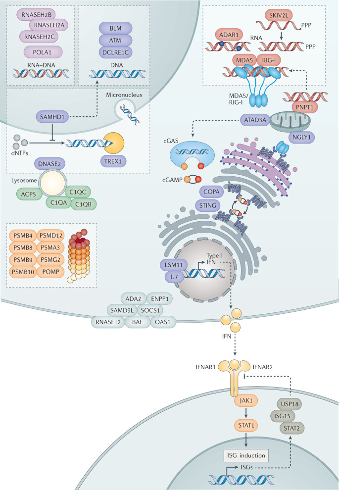
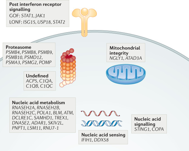
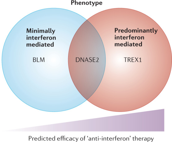
As brutally demonstrated by the COVID-19 pandemic, an effective immune system is essential for survival. Developed over evolutionary time, viral nucleic acid detection is a central pillar in the defensive armamentarium used to combat foreign microbial invasion. To ensure cellular homeostasis, such a strategy necessitates the efficient discrimination of pathogen-derived DNA and RNA from that of the host. In 2011, it was suggested that an upregulation of type I interferon signalling might serve as a defining feature of a novel set of Mendelian inborn errors of immunity, where antiviral sensors are triggered by host nucleic acids due to a failure of self versus non-self discrimination. These rare disorders have played a surprisingly significant role in informing our understanding of innate immunity and the relevance of type I interferon signalling for human health and disease. Here we consider what we have learned in this time, and how the field may develop in the future.


Conflict of interest statement

The authors declare working as consultants for Related Sciences.

Figures

Fig. 1
Fig. 1. Putative type I interferonopathy genotypes.
Mutations affecting ribonuclease H2 (RNase H2), a trimeric protein (encoded by RNASEH2A, RNASEH2B and RNASEH2C), and POLA1, the catalytic subunit of DNA polymerase-α, an essential component of the DNA-replication machinery, are suggested to result in an alteration in cytosolic levels of RNA–DNA hybrids. Mutations in BLM, ATM and DCLRE1C, encoding the RecQ–like helicase BLM, the DNA repair protein ataxia telangiectasia mutated (ATM) and the DNA double-strand break repair protein Artemis, respectively, lead to the accumulation of products of DNA damage. SAMHD1 hydrolyses deoxynucleoside triphosphates (dNTPs) and may also play a role in DNA repair, while TREX1 degrades single-stranded and double-stranded DNA molecules. DNASE2, encoding the lysosomal endonuclease DNase II (DNASE2), promotes clearance of nucleic acids generated through apoptosis and the phagocytosis of maturating erythroblast nuclei. Mutations in LSM11 and RNU7-1 result in a disturbance of histone stoichiometry, leading to sensing of nuclear DNA. ATAD3A mutations result in a leakage of mitochondrial DNA into the cytosol. All of the aforementioned mutant genotypes signal to interferon induction through cyclic GMP–AMP (cGAMP) synthase (cGAS)–STING signalling, which leads to the production of type I interferon (via activation of TANK-binding kinase 1 (TBK1)–interferon regulatory factor 3 (IRF3) and possibly NF-κB (not shown)). Gain-of-function mutations in STING lead to constitutive translocation of the protein from the endoplasmic reticulum to the Golgi apparatus, and dominant negative mutations in coatomer subunit-α (COPA), involved in retrograde Golgi apparatus to endoplasmic reticulum vesicular transport, also result in abnormal STING trafficking. MDA5 (encoded by IFIH1) and RIG-I (encoded by DDX58) normally sense exogenous viral double-stranded RNA (dsRNA); Aicardi–Goutières syndrome-related gain-of-function mutations in MDA5 lower its activation threshold to enable sensing of endogenous dsRNA species. dsRNA-specific adenosine deaminase 1 (ADAR1) deaminates adenosine to inosine, and loss-of-function mutations are proposed to result in the generation of abnormally immunogenic dsRNA species derived from Alu inverted repeats. Mutations in any of these proteins activate interferon signalling via an RNA-sensing pathway involving MAVS. The same is true of mutations in the mitochondrial ribonuclease polynucleotide phosphorylase (PNPT1) and in the RNA helicase SKIV2L, which plays a role in limiting the activation of the cytosolic dsRNA receptor machinery in response to IRE1-mediated RNA degradation. The predominant signalling pathway induced by mutations in N-glycanase (NGLY1), a conserved deglycosylation enzyme, remains unclear. The signalling pathways involved in inducing an interferon signature due to mutations in C1q, the first protein of the classical complement pathway, encoded by C1QA, C1QB and C1QC, and ACP5, which encodes the lysosomal phosphatase tartrate-resistant acid phosphatase (TRAP), and in the multiple genes encoding distinct proteasomal components, are also currently undefined. After induction, interferon (IFN) binds to heterodimeric interferon-α/β receptor 1 (IFNAR1), leading to phosphorylation of JAK1 and TYK2 and subsequent activation of the transcription factor complex ISGF3. ISGF3 binds to interferon-stimulated response elements in gene promoters and induces the expression of interferon-stimulated genes (ISGs). USP18 is a negative regulator of signalling downstream of IFNAR1. Ubiquitin-like protein ISG15 stabilizes the level of intracellular USP18. Mutations in ISG15 cause a reduction of the level of USP18, resulting in enhanced interferon production. While STAT2 plays a role in positive interferon signal induction, homozygous separation-of-function mutations at p.Arg148 lead to disruption of a role in limiting IFNAR2 signalling. Gain-of-function mutations in JAK1 and STAT2 lead to enhanced type I interferon and other cytokine signalling pathways. We are uncertain of the interferon status of disease related to mutations in adenosine deaminase 2 (ADA2), sterile α-motif domain-containing protein 9-like (SAMD9L), suppressor of cytokine signalling 1 (SOCS1), barrier-to-autointegration factor 1 (BAF), ENPP1 ectonucleotide pyrophosphatase/phosphodiesterase (ENPP1), ribonuclease T2 (RNASET2) and oligoadenylate synthetase 1 (OAS1), encoded by CECR1, SAMD9L, SOCS1, BAF1, ENPP1, RNASET2 and OAS1 respectively, either because of a current lack of or because of the presence of conflicting clinical, in vitro, ex vivo or in vivo evidence. PPP, triphosphate.
Fig. 2
Fig. 2. Categorization of putative type I interferonopathy genotypes according to the function of the encoded gene product.
Seventeen of the 38 putative type I interferonopathy mutated gene products act directly on nucleic acid substrates (nucleic acid ‘metabolism’) or are involved in nucleic acid sensing, two have a primary role in transducing a DNA signal to an innate immune response, five are involved in downstream regulation of type I interferon receptor signalling and two contribute to the maintenance of mitochondrial integrity. Eight further genes relate to the proteasome, where the precise link to type I interferon induction remains unclear. The relationship of mutations in ACP5 and C1QA, C1QB, and C1QC mutations to disease remains unproven to our mind. GOF, gain of function; LONF, loss of negative feedback.
Fig. 3
Fig. 3. Relationship between the relative contribution of interferon-mediated and non-interferon-mediated inflammation to putative type I interferonopathy phenotypes.
The effect of ‘anti-interferon’ treatments is expected to depend on the contribution of upregulated interferon signalling to the associated phenotype. Exemplar genotypes are given where the clinical efficacy of such therapies is expected to differ. Other factors (for example, the stage in the disease process at which treatment is started) will likely also be important in determining therapeutic efficacy.

References

    1. Crow YJ. Type I interferonopathies: a novel set of inborn errors of immunity. Ann. N. Y. Acad. Sci. 2011;1238:91–98. doi: 10.1111/j.1749-6632.2011.06220.x. - DOI - PubMed
    1. Crow YJ, Lebon P, Casanova JL, Gresser I. A brief historical perspective on the pathological consequences of excessive Type I interferon exposure in vivo. J. Clin. Immunol. 2018;38:694–698. doi: 10.1007/s10875-018-0543-6. - DOI - PubMed
    1. Gresser I. Can interferon induce disease? Interferon. 1982;4:95–127. - PubMed
    1. Casanova JL. Ion Gresser. J. Interferon Cytokine Res. 2019;39:317–320. doi: 10.1089/jir.2018.29015.mem. - DOI
    1. Lebon P, et al. Intrathecal synthesis of interferon-alpha in infants with progressive familial encephalopathy. J. Neurol. Sci. 1988;84:201–208. doi: 10.1016/0022-510X(88)90125-6. - DOI - PubMed

Publication types